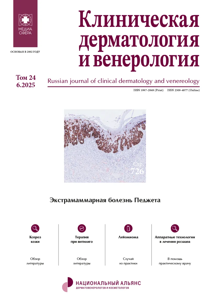
Экстрамаммарная болезнь Педжета
Журнал: Клиническая дерматология и венерология. 2025;24(6):726-733.
DOI: 10.17116/klinderma202524061726
Прочитано: 1382 раза
Резюме
Экстрамаммарная болезнь Педжета (ЭМБП) впервые описана H. Crocker в 1889 г. у пациента с поражением кожи мошонки и полового члена. Позднее было установлено, что она встречается преимущественно на участках кожи, богатых апокриновыми потовыми железами. В соответствии с классификацией ее подразделяют на первичную и вторичную, последняя связана с аденокарциномами внутренних органов. Клиническая картина заболевания неспецифична и требует дифференциальной диагностики с воспалительными заболеваниями гениталий. Определяющим методом постановки диагноза является гистологическое и иммуногистохимическое исследование. Гистологическим маркером ЭМБП является обнаружение клеток Педжета, выявляемых как при рутинной микроскопии, так и при использовании гистохимических красителей. Для дифференциальной диагностики первичной и вторичной ЭМБП используются иммуногистохимические маркеры: первичная является GCDFP-15 (+), CK7 (+) и CK20 (–), а вторичная может быть GCDFP-15 (–), CK20 (+), CK7 (±) в зависимости от характера опухоли, вызвавшей ее появление. Из-за редкой встречаемости заболевания опыт его лечения ограничен. Хирургическое иссечение является методом выбора для локальных поражений без метастазов. В последнее время накоплен опыт использования местных цитотоксических препаратов (блеомицин, 5-фторурацил) и имиквимода при лечении неинвазивной ЭМБП. Системная химиотерапия (винкристин, доцетаксел, карбоплатин, 5-фторурацил, митомицин-с, этопозид) показана при метастатической форме ЭМБП. Основными факторами, определяющими прогноз, являются глубина инвазии и наличие или отсутствие метастазов.
Ключевые слова
- экстрамаммарная болезнь Педжета
- классификация
- гистологическая диагностика
- иммуногистохимия
- обследование
- лечение
Дата поступления: 12.02.2025
Дата принятия в печать: 15.09.2025
Дата публикации: 27.12.2025
- Paget J. One disease of the mammary areola proceeding cancer of the mammary gland. St Barth Hosp Rep. 1874;10:87-89.
- Crocker HR. Paget’s disease affecting the scrotum und penis. Trans Pathol Soc Lond. 1889;40: 187-191.
- Lopes Filho LL, Lopes IM, Lopes LR, et al. Mammary and extramammary Paget’s disease. An. Bras. Dermatol. 2015;90(2):225-231. https://doi.org/10.1590/abd1806-4841.20153189
- Claire KS, Hoover A, Ashack K, Khachemoune A. Extramammary Paget’s disease. Dermatology Online Journal. 2019;25(4):1-11. https://doi.org/10.5070/D3254043591
- McDaniel B, Brown F, Crane JS. Extramammary Paget Disease. [Updated 2023 Jul 31]. In: StatPearls [Internet]. Treasure Island (FL): StatPearls Publishing; 2024 Jan. Accessed June 06, 2024. https://www.ncbi.nlm.nih.gov/books/NBK493224/
- Cheng PS, Lu CL, Cheng CL, Lai FJ. Significant male predisposition in extramammary Paget disease: a nationwide population-based study in Taiwan. Br J Dermatol. 2014;171(1):191-193. [PMID: 24471479].
- Chanda JJ. Extramammary Paget’s disease: prognosis and relationship to internal malignancy. J Am Acad Dermatol. 1985 Dec;13(6):1009-1014. PMID: 3001158. https://doi.org/10.1016/s0190-9622(85)70254-x
- Scarbrough CA, Vrable A, Carr DR. Definition, Association with Malignancy, Biologic Behavior, and Treatment of Ectopic Extramammary Paget’s Disease: A Review of the Literature. The Journal of Clinical and Aesthetic Dermatology. 2019 Aug;12(8):40-44.
- WHO Classification of Skin Tumours. DE Elder, D Massi, RA Scolyer, et al. 4th ed. Lyon, 2018;217-218.
- Kyriazanos ID, Stamos NP, Miliadis L, Noussis G, Stoidis CN. Extra-mammary Paget’s disease of the perianal region: a review of the literature emphasizing the operative management technique. Surg Oncol. 2011 Jun;20(2): e61-71. PMID: 20884199. https://doi.org/10.1016/j.suronc.2010.09.005
- Lloyd J, Flanagan AM. Mammary and extramammary Paget’s disease. J Clin Pathol. 2000 Oct;53(10):742-749. PMID: 11064666; PMCID: PMC1731095. https://doi.org/10.1136/jcp.53.10.742
- Lagios MD, Westdahl PR, Rose MR, Concannon S. Paget’s disease of the nipple. Alternative management in cases without or with minimal extent of underlying breast carcinoma. Cancer. 1984;54:545-551.
- Jahn HI, Osther PJ, Nielsen EH, Rasmussen G, Andersen J. An electron microscopic study of clinical Paget’s disease of the nipple. APMIS. 1995 Sep;103:628-634.
- Willman JH, Golitz LE, Fitzpatrick JE. Vulvar clear cells of Toker: precursors of extramammary Paget’s disease. Am J Dermatopathol. 2005;27(3):185-188. PMID: 15900119.
- Belousova IE, Kazakov DV, Michal M, Suster S. Vulvar Toker Cells: The Long-Awaited Missing Link: A Proposal for an Origin-Based Histogenetic Classification of Extramammary Paget Disease. The American Journal of Dermatopathology. 2006 Feb;28(1):84-86. https://doi.org/10.1097/01.dad.0000194052.65695.f1
- Ohara K, Fujisawa Y, Yoshino K, Kiyohara Y, Kadono T, Murata Y, Uhara H, Hatta N, Uchi H, Matsushita S, Takenouchi T, Hayashi T, Yoshimura K, Fujimoto M. A proposal for a TNM staging system for extramammary Paget disease: Retrospective analysis of 301 patients with invasive primary tumors. J Dermatol Sci. 2016 Sep;83(3):234-239. Epub 2016 Jun 03. PMID: 27329007. https://doi.org/10.1016/j.jdermsci.2016.06.004
- Fernandez-Flores A, Eraña I, Cuevas J. “Extramammary-Type” Paget Disease of the Breast. Am J Dermatopathol. 2018 Oct; 40(10):711-720. PMID: 30234560. https://doi.org/10.1097/DAD.0000000000001105
- Căruntu C, Zurac SA, Jugulete G, Boda D. Extramammary Paget’s disease in an HIV-positive patient. Rom J Morphol Embryol. 2017;58(3):1009-1015. PMID: 29250682.
- Simonds RM, Segal RJ, Sharma A. Extramammary Paget’s disease: a review of the literature. Int J Dermatol. 2019 Aug;58(8):871-879. Epub 2018 Dec 19. PMID: 30569580. https://doi.org/10.1111/ijd.14328
- Inui S, Fukuhara S, Asada H, et al. Double involvement of extramammary Paget’s disease in the genitalia and axilla. J Dermatol. 2000;27:409-412. https://doi.org/10.1111/j.1346-8138.2000.tb02194.x
- Murata Y, Kumano K, Tan M. Underpants-pattern erythema: a previously unrecognized cutaneous manifestation of extramammary Paget’s disease of the genitalia with advanced metastatic spread. J Am Acad Dermatol. 1999; 40:949-956. https://doi.org/10.1016/s0190-9622(99)70083-6
- Smirnova IO, Petunova YG, Belousova IE, Konstantinova AM, Shelekhova KV, Oganesyan MV. Difficulties of the diagnosis of the extra mammary Paget disease. Russian Journal of Clinical Dermatology and Venereology. 2018;17(3):36-40. https://doi.org/10.17116/klinderma201817336
- Konstantinova AM, Shelekhova KV, Michal M, Kazakov DV. Primary extramammary Paget disease of the anogenital area: morphological features of the tumor and their importance for the disease recurrence. Sibirskiy nauchnyy meditsinskiy zhurnal = Siberian Scientific Medical Journal. 2021;41(1):50-59. (In Russ.). https://doi.org/10.18699/SSMJ20210105
- Lam C, Funaro D. Extramammary Paget’s disease: Summary of current knowledge. Dermatol Clin. 2010;28(4):807-926. PMID: 20883922.
- Ichiyama T, Gomi D, Fukushima T, Kobayashi T, Sekiguchi N, Sakamoto A, Sasaki S, Mamiya K, Koizumi T, Hama Y. Successful and long-term response to trastuzumab plus paclitaxel combination therapy in human epidermal growth factor receptor 2-positive extramammary Paget’s disease: A case report and review of the literature. Mol Clin Oncol. 2017;7(5):763-766. PMID: 29181166.
- Sekiguchi N, Kubota S, Noguchi T, Fukushima T, Kobayashi T, Kanda S, Koizumi T, Miyake T, Shirai T, Okuyama R. Experiences of trastuzumab plus paclitaxel combination therapy in metastatic human epidermal growth factor receptor 2-positive extramammary Paget’s disease: Four cases and a review. J Dermatol. 2020 Nov;47(11):1276-1279. Epub 2020 Jul 24. PMID: 32706146. https://doi.org/10.1111/1346-8138.15515
- Shin DS, Sherry T, Kallen ME, Wong S, Drakaki A. Human Epidermal Growth Factor Receptor 2 (HER-2/neu)-Directed Therapy for Rare Metastatic Epithelial Tumors with HER-2 Amplification. Case Rep Oncol. 2016 Jun 11;9(2):298-304. PMID: 27403128; PMCID: PMC4924467. https://doi.org/10.1159/000445827
- Hata M, Koike I, Wada H, Miyagi E, Kasuya T, Kaizu H, Mukai Y, Inoue T Postoperative radiation therapy for extramammary Paget’s disease. Br J Dermatol. 2015;172(4):1014-1020. PMID: 25139574.
- Morris CR, Hurst EA. Extramammary Paget’s Disease: A Review of the Literature. Part II: Treatment and Prognosis. Dermatol Surg. 2020;46(3):305-311. https://doi.org/10.1097/DSS.0000000000002240
- Wollina U, Goldman A, Bieneck A, Abdel-Naser MB, Petersen S. Surgical Treatment for Extramammary Paget’s Disease. Curr Treat Options Oncol. 2018;19(6):27. https://doi.org/10.1007/s11864-018-0545-x
- Padrnos L, Karlin N, Halfdanarson TR. Mayo Clinic Cancer Center Experience of Metastatic Extramammary Paget Disease 1998-2012. Rare Tumors. 2016;8(4):6804. https://doi.org/10.4081/rt.2016.6804
- Preti M, Micheletti L, Borella F, Cosma S, Marrazzu A, Gallio N, et al. Vulvar Paget’s disease and stromal invasion: Clinico-pathological features and survival outcomes. Surg Oncol. 2021;38:101581. https://doi.org/10.1016/j.suronc.2021.101581
- Coldiron BM, Goldsmith BA, Robinson JK. Surgical treatment of extramammary Paget’s disease. A report of six cases and a reexamination of Mohs micrographic surgery compared with conventional surgical excision. Cancer. 1991 Feb 15;67(4):933-938. PMID: 1846769. https://doi.org/10.1002/1097-0142(19910215)67:4<933::aid-cncr2820670413>3.0.co;2-3
- Córdoba A, Iglesias ME, Rodríguez I, Yanguas JI. Extramammary Paget’s disease with frontotemporal involvement: a case report and review of the literature. Actas Dermosiffliogr. 2013;104(4):355-357. Epub 2013 Apr 06. PMID: 23567449. https://doi.org/10.1016/j.adengl.2013.03.006
- Youssef AA, Reygagne P. Successful treatment of extramammary Paget’s disease of the scalp with photodynamic therapy. Int J Dermatol. 2016;55(5): 580-582.
- Kariya K, Tsuji T, Schwartz RA. Trial of low-dose 5- fluorouracil/cisplatin therapy for advanced extramammary Paget’s disease. Dermatol Surg. 2004;30(2 Pt 2):341-344. [PMID: 14871231].
- Guercio BJ, Iyer G, Kidwai WZ, Lacouture ME, Ghafoor S, Rossi AM, et al. Treatment of Metastatic Extramammary Paget Disease with Combination Ipilimumab and Nivolumab: A Case Report. Case Rep Oncol. 2021 Mar 12; 14(1):430-438. PMID: 33790764; PMCID: PMC7983595. https://doi.org/10.1159/000514345
- Emelyanova OO, Zikirjahodzhaev AD, Saribekyan EK, Filonenko EV. The current consensus for the diagnostic and treatment of extramammary Paget’s disease. Vestnik Dermatologii i Venerologii. 2023;99(3):23-32. https://doi.org/10.25208/vdv1400
- Yao H, Xie M, Fu S, et al. Survival analysis of patients with invasive extramammary Paget disease: implications of anatomic sites. BMC Cancer. 2018; 18(1):403. Published 2018 Apr 10. https://doi.org/10.1186/s12885-018-4257-1
- Hatta N, Yamada M, Hirano T, Fujimoto A, Morita R. Extramammary Paget’s disease: treatment, prognostic factors and outcome in 76 patients. Br J Dermatol. 2008 Feb;158(2):313-318. Epub 2007 Nov 19. PMID: 18028492. https://doi.org/10.1111/j.1365-2133.2007.08314.x


Рекомендуем статьи по данной теме:
Эффективность комплексного лечения пациенток с острым и обострением хронического сальпингоофорита с применением биологического лекарственного препарата секретома лейкоцитов периферической крови.Проблемы репродукции. 2026;32(4):91-101
Современные подходы к терапии NCMT-инфекций, передаваемых половым путем (N. gonorrhoeae, C. trachomatis, M. genitalium, T. vaginalis). Часть II. M. genitalium.Клиническая дерматология и венерология. 2026;25(4):603-612
Наследственная атрофия зрительных нервов Лебера.Вестник офтальмологии. 2026;142(4):105-111
О классификации автомобильной травмы.Судебно-медицинская экспертиза. 2026;69(4):19-23
Муцинозная аденокарцинома червеобразного отростка, имитирующая опухоль яичника.Архив патологии. 2026;88(4):55-59
Характеристика субпопуляций лимфоцитов и клеток системы мононуклеарных фагоцитов печени в острый период и в отдаленные сроки после перенесенного заболевания COVID-19.Архив патологии. 2026;88(4):5-13
Химические ожоги у детей.Хирургия. Журнал им. Н.И. Пирогова. 2026;(7):104-110
Клинический случай диагностики и лечения экстраносоглоточной ангиофибромы.Вестник оториноларингологии. 2026;91(3):111-114
Инфекционный уретрит у мужчин: этиология, эпидемиология, диагностика и лечение (аналитический обзор).Клиническая дерматология и венерология. 2026;25(3):464-472
Синдром эмоционального выгорания: профилактика, лечение и реабилитация.Восстановительные биотехнологии, профилактическая, цифровая и предиктивная медицина. 2025;2(3):61-68