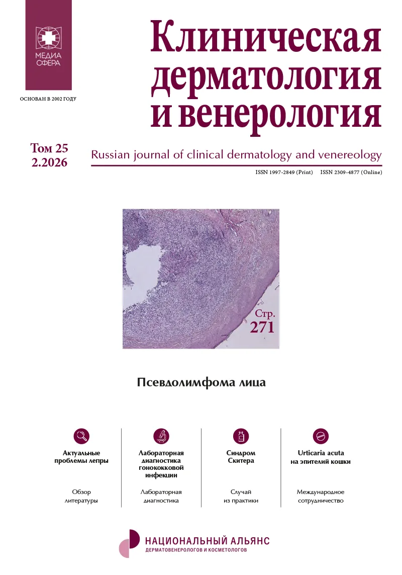
Случай псевдолимфомы кожи лица как реакция на профессиональное воздействие дихлорэтана
Журнал: Клиническая дерматология и венерология. 2026;25(2):271-276.
DOI: 10.17116/klinderma202625021271
Прочитано: 772 раза
Резюме
Доброкачественная лимфоплазия кожи — наиболее часто встречающаяся клиническая форма псевдолимфомы кожи, неспецифичность клинических проявлений которой создает трудности для диагностики, приводит к ошибочным диагнозам. Представлен клинический случай развития доброкачественной лимфоплазии кожи лица у пациента 53 лет, обратившегося к дерматовенерологу с жалобой на появление множественных плотных зудящих узелков на коже лица. Первичный клинический диагноз: акнеформный дерматоз? Пациент направлен на консультацию в ГБУ СО УрНИИДВиИ. Из анамнеза установлено: около 4 лет имеет профессиональный контакт с высокоопасным химическим веществом 1,2-дихлорэтаном, оказывающим кожно-резорбтивное действие. Предварительный клинический диагноз: фотодерматоз? саркоидоз кожи? Проведенная инцизионная биопсия кожи в области патологического процесса выявила атипичную лимфоидную пролиферацию, нодулярный неэпидермотропный инфильтрат преимущественно из мелких лимфоидных клеток с примесью гистиоцитов, единичных эозинофилов. Для исключения злокачественного лимфопролиферативного процесса проведено иммуногистохимическое исследование. Заключение: инфильтрат имеет смешанный, преимущественно T-клеточный состав. Гистоархитектоника и иммунофенотип инфильтрата характерны для доброкачественной лимфоплазии кожи (нодулярной T-клеточной псевдолимфомы). На основании клинической картины, данных иммуногистохимического и патоморфологического исследований биоптата кожи выставлен диагноз: T-клеточная псевдолимфома кожи, инфильтративно-бляшечная форма доброкачественной лимфоплазии кожи Jessner—Kanoff’s. После проведенной терапии при исключении контакта с дихлорэтаном положительная динамика кожного процесса. Клиническое наблюдение подтверждает необходимость детального изучения анамнеза заболевания (включая профессиональный маршрут), что позволяет идентифицировать провоцирующий фактор развития заболевания, провести дифференциальную диагностику на основе анализа данных анамнеза, клиники, патоморфологии, иммуногистохимии.
Ключевые слова
- псевдолимфома кожи
- клинический случай
- дихлорэтан
- дифференциальная диагностика
- патоморфология
- иммуногистохимия
Дата поступления: 24.09.2025
Дата принятия в печать: 09.01.2026
Дата публикации: 30.04.2026
- Олисова О.Ю., Потекаев Н.С. Псевдолимфомы кожи. М.: Практика; 2013.
- Олисова О.Ю., Снарская Е.С., Теплюк Н.П., Варшавский В.А., Золотарева Е.С., Махмуди В. К вопросу диагностики и лечения псевдолимфомы кожи на примере клинического случая. Российский журнал кожных и венерических болезней. 2023;26(2):105-118. https://doi.org/10.17816/dv217710
- Олисова О.Ю., Анпилогова Е.М., Косумова Х.С., Никулина Е.Е. Совершенствование диагностики и лечения доброкачественной лимфоплазии кожи. Российский журнал кожных и венерических болезней. 2021;24(5):429-442. https://doi.org/10.17816/dv96711
- Потекаев Н.С., Олисова О.Ю., Галил-Оглы Г.А. Диагностика истинной псевдолимфомы кожи. Международный медицинский журнал. 2003;2:117-120.
- Kempf W, Mitteldorf C, Cerroni L, Willemze R, Berti E, Guenova E, Scarisbrick J, Battistella M. Classifications of cutaneous lymphomas and lymphoproliferative disorders: An update from the EORTC cutaneous lymphoma histopathology group. J Eur Acad Dermatol Venereol. 2024;38(8):1491-1503. https://doi.org/10.1111/jdv.19987
- Олисова О.Ю., Теплюк Н.П., Гаранян Л.Г., Пятилова П.М. Доброкачественная лимфоплазия кожи, развившаяся на месте татуировки. Российский журнал кожных и венерических болезней. 2015;18(2):23-26.
- Тарасенко Г.Н., Бобин А.Н., Кузьмина Ю.В., Овсянникова М.Р., Тарасенко Ю.Г. Случай псевдолимфомы кожи: как реакция на вакцинацию. Госпитальная медицина: наука и практика. 2022;6:49-52.
- Олисова О.Ю., Анпилогова Е.М. Новые возможности в лечении доброкачественной лимфоплазии кожи. Российский журнал кожных и венерических болезней. 2020;23(1):17-22. https://doi.org/10.17816/dv2020117-22
- Потекаев Н.С., Сергеев Ю.В. Псевдолимфомы. В кн. Кожные и венерические болезни. Под ред. Ю.К. Скрипкина. Том 3. М.: Медицина; 1996:106-112.
- Снарская Е.С., Гришина В.Б., Гаранян Л.Г. Лимфоцитарная инфильтрация Иесснера—Канофа. Российский журнал кожных и венерических заболеваний. 2015;18(4):17-20.


Рекомендуем статьи по данной теме:
Флотирующее образование правого предсердия у пациентки с вероятной хронической тромбоэмболической легочной гипертензией.Хирургия. Журнал им. Н.И. Пирогова. 2026;(9-2):148-154
Клинико-морфологические прогностические показатели рецидива полипозного риносинусита.Российская ринология. 2026;34(3):40-46
Случай болезни Гровера.Клиническая дерматология и венерология. 2026;25(4):557-560
Эндометриоидная аденокарцинома эндометрия IA стадии, ассоциированная с беременностью: возможности отсрочки лечения (клиническое наблюдение и обзор литературы).Онкология. Журнал им. П.А. Герцена. 2026;15(4):76-81
Муцинозная аденокарцинома червеобразного отростка, имитирующая опухоль яичника.Архив патологии. 2026;88(4):55-59
Характеристика субпопуляций лимфоцитов и клеток системы мононуклеарных фагоцитов печени в острый период и в отдаленные сроки после перенесенного заболевания COVID-19.Архив патологии. 2026;88(4):5-13
Морфологическая и иммуногистохимическая характеристика сердца пациентов с COVID-19.Архив патологии. 2026;88(1):44-51
Особенности дифференциальной диагностики онкологической патологии на амбулаторном этапе.Профилактическая медицина. 2025;28(2):93-95
Клинический случай ВИЧ-ассоциированной формы саркомы Капоши.Клиническая дерматология и венерология. 2024;23(6):782-786
Гигантский перикраниальный синус с образованием обширного дефекта кости свода черепа затылочной области. Клинический случай и обзор литературы.Вопросы нейрохирургии имени Н.Н. Бурденко. 2024;88(6):77-87