- Media Sphera
- Publishers





Introduction
The odontogenic keratocyst (OKC) was established as a separate pathologic entity in 1956 by philipsen and this developmental entity makes up about 11.2% of all the odontogenic cysts [1]. OKC is unique among the other odontogenic cysts because of its distinct histologic characteristics and high recurrence rate [2—4]. The clinical aggressiveness and recurrence of OKCs have been a topic of study in literature previously [5, 6]. The presence of non-keratinized odontogenic epithelial lining, and its intense proliferative activity to form rete ridges and occasional arcading patterns are the main histological features of inflamed OKCs, which makes them indistinguishable from the other odontogenic cysts which are secondarily infected [7].
The proliferative potential of odontogenic keratocystic lining secondary to inflammation and the influence of this inherent growth potential of this epithelial lining on the recurrence rate of OKC were also reported previously in literature [8—10]. The tendency of the epithelial cells to lose its characteristic features upon secondary inflammation and an increase in the expression of ki-67 and PCNA labeling index in metaplastic epithelium in areas with moderate to severe inflammation were studied previously by Benn et al in 1996 and Kaplan et al in 2004 [1, 11]. In addition, Smith g et al found that a substantial number of chronic inflammatory cell infiltrate, particularly the mast cells in the subepithelial connective tissue wall can play an important role in cyst enlargement [12]. Several immunohistochemical studies also showed an intricate relation between the inflammation and epithelial proliferation in OKC and thereby correlating the inflammation and clinical aggressiveness [13—16]. The aim of the present study is to retrospectively evaluate the association of inflammatory cell infiltrate and clinical aggressiveness of odontogenic keratocyst.
Materials and methods
This study was conducted with approval from the institutional ethics committee and included n=45 cases of OKC reported to the department of oral and maxillofacial pathology, blinded for peer review. The demographic and clinical details of the selected cases were retrieved from the histopathology reports and common clinical database. Cases with questionable and nonspecific histopathological diagnosis were excluded and only histopathologically confirmed cases of OKC were included in the present study. Slides of the selected cases were retrieved from the oral pathology department archives. Histopathological analysis of inflammation in OKC was done by two independent pathologists and the details regarding the clinical measurement of the lesions were obtained from the common clinical database.
The various criterias used to assess the clinical size of OKC and the histopathological assessment of inflammation is given in table 1 (Table 1). Based on these criterias, 45 cases of OKC were analysed histopathologically for inflammatory cell infiltrate. All the demographic, clinical, histopathological details were tabulated in Microsoft Excel and later exported into IBM SPSS statistics software. Pearson Chi-square test was done to analyse the association between the inflammation and clinical aggressiveness and p values less than 0.05 is considered as statistically significant.
Table 1. Showing the criteria of assessing the clinical aggressiveness and the histopathological evaluation of inflammation in OKC
| Clinical aggressiveness | Small <2cm |
|---|---|
| Moderate 2—5 cm | |
| Large >5 cm | |
| Type of inflammatory cell infiltrate | Acute |
| Chronic | |
| Amount of inflammatory cell infiltrate | Mild |
| Moderate | |
| Severe | |
| Distribution of inflammatory cell infiltrate | Localised |
| Diffused |
Results
71.1% of the OKC cases selected showed chronic inflammatory cell infiltrate followed by mixed (24.4%). Moderate to severe inflammation was seen in 71.1% of the cases and Diffused pattern of distribution (64.4%) of inflammatory cells is more commonly seen than localized. On assessing the clinical aggressiveness, moderately aggressive lesions were more common (Table 2).
Table 2. Showing the percentage of the type, amount and distribution of inflammatory cell infiltrate and the clinical aggressiveness of the OKC cases included in the study
| Inflammatory cell infiltrate — Type, % | Acute | 4.4 |
|---|---|---|
| Chronic | 71.1 | |
| Mixed | 24.4 | |
| Inflammatory cell infiltrate — Amount, % | Mild | 28.9 |
| Moderate | 53.3 | |
| Severe | 17.8 | |
| Inflammatory cell infiltrate — Distribution, % | Localised | 35.6 |
| Diffused | 64.4 | |
| Clinical Aggressiveness, % | Small | 31.1 |
| Moderate | 42.2 | |
| Large | 26.7 |
Among all the cases, 45% of the cases in which the epithelial lining lost its characteristic appearance and became proliferative and non-keratinized. In 88.8% of the cases with hyperplastic epithelial lining showed moderate to severe inflammatory cell infiltrate in connective tissue wall (p=0.05). The association between the clinical aggressiveness and type of inflammatory cell infiltrate was found to be statistically significant (p=0.019) (Table 3) and the OKCs with a clinical aggressive course showed more of a chronic to mixed inflammatory cell infiltration rather than acute inflammation (Fig. 1). The amount of inflammatory cell infiltrate and the clinical aggressiveness also showed statistically significant association (p=0.023) (Fig. 2).
Table 3. Showing the association between clinical aggressiveness and inflammation in OKC
| Variables | p-value (Chi-square test) | |
|---|---|---|
| Inflammatory cell infiltrate — Type | Clinical Aggressiveness | *.019 |
| Inflammatory cell infiltrate — Amount | Clinical Aggressiveness | *.023 |
| Inflammatory cell infiltrate — Amount | Epithelial lining morphology | 0.05 |
| Inflammatory cell infiltrate — Distribution | Clinical Aggressiveness | 0.206 |
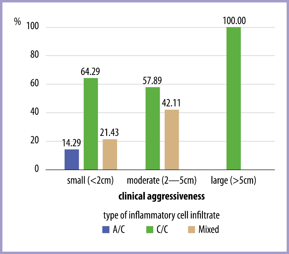
Fig. 1. Bar graph showing the association of type of inflammatory cell infiltrate and clinical aggressiveness. Chi-square test-p value = 0.019. Chronic to mixed inflammatory cell infiltrate were more associated with clinically aggressive OKCs.
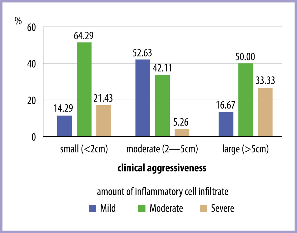
Fig. 2. Bar graph showing the association of amount of inflammatory cell infiltrate and clinical aggressiveness. Chi-square test-p value = 0.023. Moderate to severe inflammation is predominantly seen in clinically aggressive OKCs.
Discussion
OKC is a developmental cyst originating from odontogenic epithelium and accounts for 11.2% of all the odontogenic cysts [17]. Philipsen in 1956 proposed OKC as a separate pathologic entity [18]. In WHO 2005 classification of head and neck tumours, OKC has been reclassified and renamed as keratocystic odontogenic tumor (KCOT) because of its increased clinical aggressiveness and high recurrence rates [19]. Later in 2017, WHO classified the keratocystic odontogenic tumor back into the cyst category as OKC [20].
In an uninflamed OKC, the odontogenic epithelial lining is parakeratinized and corrugated with a uniform thickness of 6—10 cell layers and a polarised basal cell layer [21]. The clinical aggressiveness and high recurrence rate of odontogenic keratocyst is due to the presence of satellite cysts, daughter cysts and the flat connective tissue epithelial interface which makes it difficult to remove during surgical enucleation [1]. Thus the remaining fragmented epithelial lining following an improper enucleation procedure by the surgeon can lead to the formation of new cysts [22]. This also suggests the importance of modulation of the proliferation capacity of odontogenic epithelial lining cells and its impact on rate of recurrence and clinical aggressiveness [18].
Even though the OKC is a developmental cyst, most of the reported cases in the literature were inflamed OKC [23—25]. In the present study also, 71.1% of the cases showed chronic inflammatory cells in their connective tissue walls which was in line with the previous literature. Several studies have also found a direct influence of inflammatory cell infiltrate on the epithelial cell proliferation. This inflammation induced hyper proliferative activity of cells o OKC lining correlates with the increased activity of the cystic lining and clinical aggressiveness [1].
The association between the inflammation and proliferation of epithelial lining cells in OKC can be direct and indirect. The direct mechanism is by the adhesion of inflammatory cells to the epithelial lining and the indirect mechanism is by the response of the epithelial cells to a series of chemokines secreted by inflammatory cells [1]. The present study was also in accordance with the same because in 45% of the cases the epithelial lining lost its characteristic appearance and became proliferative and non-keratinized and 88.8% of the cases with hyperplastic epithelial lining showed moderate to severe inflammatory cell infiltrate in their connective tissue wall (p=0.05). This indicates that there could be a direct or indirect association between the inflammation in the connective tissue wall, proliferation capacity of epithelial lining and its morphology. The above mentioned observation in the present study is supported by the observation of Paula et al, in which they established that the positive cell count of cell proliferation markers such as PCNA and Ki-67 is significantly higher in inflamed OKC compared with non-inflamed OKC (16). Apart from Paula et al., Ninomiya et al. and Nakamura et al. also studied expression of Ki 67 in odontogenic cysts [26, 27] but the disparity in thickness of epithelial lining was not taken into account in their studies. However Kaplan et al studied the expression of this proliferation marker in different fields of OKC and found that the inflammation adjacent to the epithelial cyst lining had a localized effect on epithelium which might induce proliferation in the epithelial lining and a focal increase in expression of Ki-67 but not PCNA [1]. However, this association has not been investigated thoroughly in the previous studies and the current study also gives a preliminary light in this area.
The present study also showed a statistically significant relation between the amount of inflammatory cell infiltrate and the clinical aggressiveness (Fig. 3). Among the cases included in the study, 83.3% of the clinically large lesions or clinically aggressive OKCs showed moderate to severe inflammatory cell infiltrate in their connective tissue wall (Fig. 4). This could be due to the induction of epithelial proliferation by intense inflammatory reaction and thereby increasing its aggressiveness. The chronic inflammatory cells like plasma cells, lymphocytes and macrophages were seen associated with more aggressive lesions compared to less aggressive lesions (Fig. 5). This could be explained by production and secretion of growth factors and cytokines such as interleukin-1, interleukin-6 and tumor necrosis factor (TNF) by the chronic inflammatory cells [28—30]. Apart from this, Li et al suggested that the epidermal growth factor expression by odontogenic epithelial lining is related to the presence of inflammation within the connective tissue wall [31]. Even though the present study showed a statistically significant association, the cytokines can have a varied impact on epithelial lining, it can have a positive and negative impact on cell proliferation, which cannot be assessed by the current study. Hence, an extensive study connecting the molecular mechanisms are needed to prove this association in a more scientific form.
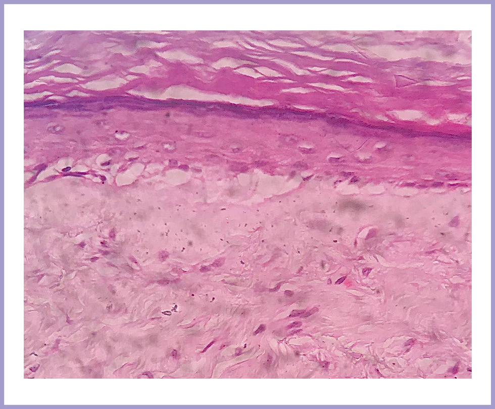
Fig. 3. Orthokeratinized OKC with mild inflammatory cells infiltrate in the connective tissue wall.
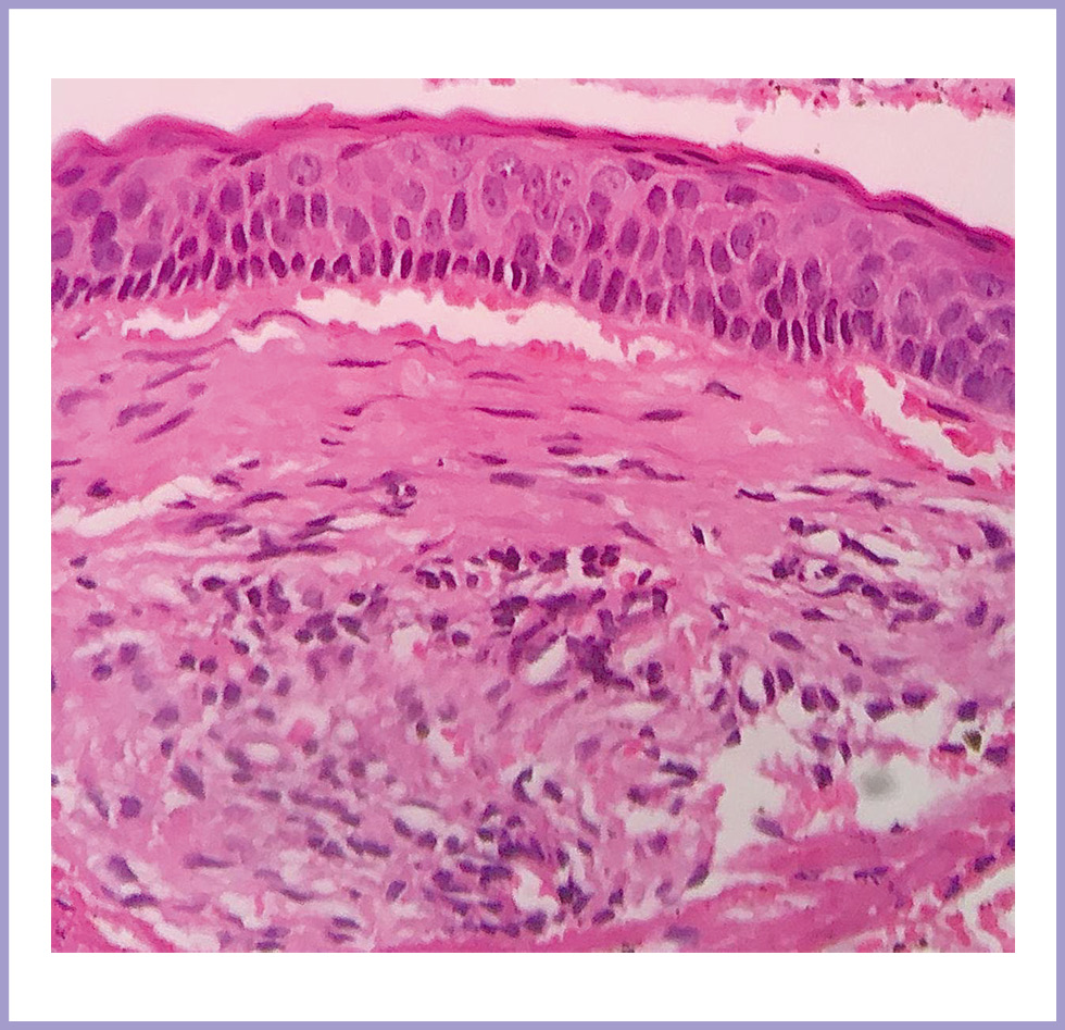
Fig. 4. OKC with moderate chronic inflammatory cells infiltrate in the connective tissue wall and detachment of the epithelial lining from connective tissue wall.
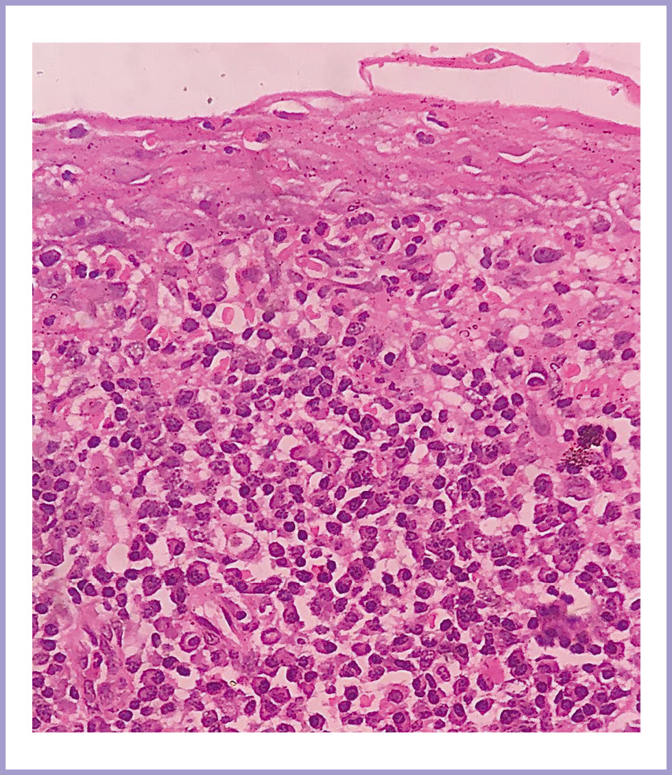
Fig. 5. OKC with intense chronic inflammatory cells infiltrate in the connective tissue wall. The epithelial lining lost its characteristic parakeratin corrugated surface layer and palisaded and columnar basal cell layer.
Lack of recording of inflammation in field specific manner and the less sample size are the main drawbacks of the present study. Immunohistochemical and molecular level studies could better aid in establishing the significant association between the inflammation and clinical aggressiveness and which can further help in the betterment of prognosis of OKC.
Conclusion
However, the present study could establish a statistically significant association between clinical aggressiveness with type and amount of inflammatory cell infiltrate, more futuristic studies with increased sample size, Immunohistochemical and molecular level techniques should be conducted to establish the association.
The authors declare no conflict of interest.