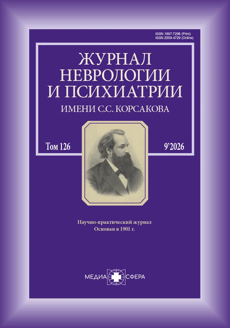
Астроциты как центральный структурно-функциональный компонент развития смешанной деменции
Журнал: Журнал неврологии и психиатрии им. С.С. Корсакова. 2026;126(9):7-14.
DOI: 10.17116/jnevro20261260917
Прочитано: 93 раза
Резюме
Смешанная деменция, характеризующаяся сочетанием патологии, вызванной болезнью Альцгеймера и цереброваскулярным заболеванием (ЦВЗ), широко распространена в пожилом возрасте. Накопленные данные свидетельствуют о том, что ЦВЗ является активным участником нейродегенеративного процесса: она потенцирует амилоидный и тау-каскады и усиливает прогрессирующую дезинтеграцию функциональных и структурных сетей мозга. В обзоре представлены данные, свидетельствующие о том, что астроциты являются центральным интегративным элементом, связывающим патологию бета-амилоида и тау-белка, дисфункцию нейроваскулярной единицы, нарушения гематоэнцефалического барьера и изменения глимфатической системы при смешанной деменции. Обсуждаются гомеостатические функции астроцитов и их реактивная трансформация с усилением передачи сигналов Ca2+, метаболическое взаимодействие, поляризация аквапорина-4 (AQP4) и фенотипы воспаления. Астроциты представляют собой ключевые модуляторы системной организации мозговой активности и центральное звено конвергенции патологических процессов при смешанной деменции. Таргетное воздействие на астроцит-опосредованные механизмы может открыть новые терапевтические возможности для коррекции бета-амилоид- и тау-протеинопатии.
Ключевые слова
- астроциты
- смешанная деменция
- болезнь Альцгеймера
- сосудистые когнитивные нарушения
- нейроваскулярная единица
- глимфатическая система
- гематоэнцефалический барьер
- кислый глиофибириллярный белок
- тау-белок
Дата поступления: 31.03.2026
Дата принятия в печать: 24.05.2026
Дата публикации: 20.10.2026
- Saito S, Ihara M, Yamaguchi Y. Astrocyte-vascular interplay in Alzheimer’s disease pathology. Int J Mol Sci. 2023;24(4):3751. https://doi.org/10.3390/ijms24043751
- Захаров В.В., Черноусов П.А., Вехова К.А. и др. Когнитивные нарушения у пациентов с артериальной гипертензией. Журнал неврологии и психиатрии им. С.С. Корсакова. 2024;124(4-2):41-48. https://doi.org/10.17116/jnevro202412404241
- Боголепова А.Н. Сосудистые когнитивные нарушения. Журнал неврологии и психиатрии им. С.С. Корсакова. 2022;122(10):17-23. https://doi.org/10.17116/jnevro202212210117
- Левин О.С., Боголепова А.Н., Лобзин В.Ю. Общие механизмы патогенеза нейроденеративных и цереброваскулярных заболеваний и возможности их коррекции. Журнал неврологии и психиатрии им. С.С. Корсакова. 2022;122(5):11-16. https://doi.org/10.17116/jnevro202212205111
- Nichols E, Merrick R, Hay S, et al. Prevalence, correlation, and co-occurrence of neuropathologies in autopsied older adults. Alzheimers Res Ther. 2023;15(1):123-132. https://doi.org/10.1016/S2666-7568(23)00019-3
- Jellinger KA, Attems J. Neuropathological evaluation of mixed dementia: Alzheimer’s disease and vascular brain injury. Acta Neuropathol. 2021;141(1):1-22.
- Burgueño-García I, López-Martínez M, Uceda-Heras A, et al. Neuropathological heterogeneity of dementia due to mixed Alzheimer and vascular pathology. J Clin Med. 2024;13(22):6755. https://doi.org/10.3390/jcm13226755
- Zlokovic BV. Neurovascular dysfunction and faulty amyloid β-peptide clearance in Alzheimer disease. Nat Rev Neurosci. 2012;13(10):723-738. https://doi.org/10.1038/nrn3268
- Eisenmenger LB, Peret A, Famakin BM, et al. Vascular contributions to Alzheimer’s disease: dysregulation of neurovascular coupling, BBB disruption and Aβ clearance. Transl Res. 2022;254:41-53. https://doi.org/10.1016/j.trsl.2022.12.003
- Iadecola C. Cerebral blood flow regulation and neurovascular dysfunction in Alzheimer disease. Nat Rev Neurosci. 2017;18(4):251-262. https://doi.org/10.1038/nrn.2017.60
- Sweeney MD, Montagne A, Sagare AP, et al. Microvascular contributions to Alzheimer disease pathogenesis: endotheliopathy and vascular dysfunction. J Clin Invest. 2023;133(3):e159453
- Ang PS, Zhang DM, Azizi SA, et al. The glymphatic system and cerebral small vessel disease: implications for waste clearance and neurodegeneration. J Stroke Cerebrovasc Dis. 2024;33(3):107557. https://doi.org/10.1016/j.jstrokecerebrovasdis.2024.107557
- Bommarito G, Griffa A, Michel P, et al. Cerebral small vessel disease in memory center patients with dementia with Lewy bodies and Alzheimer’s disease. Alzheimers Res Ther. 2025;17(1):154-159. https://doi.org/10.1186/s13195-025-01805-8
- Verkhratsky A, Nedergaard M. Physiology of astroglia. Physiol Rev. 2018;98(1):239-389. https://doi.org/10.1152/physrev.00042.2016
- Filosa JA, Morrison HW, Iddings J, et al. Beyond neurovascular coupling, role of astrocytes in the regulation of vascular tone. Neurosci. 2016;323:96-109. https://doi.org/10.1016/j.neuroscience.2015.03.064
- Kugler EC, Greenwood J, MacDonald RB. The «Neuro-Glial-Vascular» unit: the role of glia in neurovascular unit formation and dysfunction. Front Cell Dev Biol. 2021;9:732820. https://doi.org/10.3389/fcell.2021.732820
- Mishra A, Reynolds JP, Chen Y, et al. Astrocytes mediate neurovascular signaling to capillary pericytes but not to arterioles. Nat Neurosci. 2016;19(12):1619-1627. https://doi.org/10.1038/nn.4428
- Федин А.И. Глимфатическая система мозга — нейробиология и клиническая патология. Журнал неврологии и психиатрии им. С.С. Корсакова. 2023;123(5):13-19. https://doi.org/10.17116/jnevro202312305113
- Price BR, Norris C, Sompol P, et al. An emerging role of astrocytes in vascular contributions to cognitive impairment and dementia (VCID). J Neurochem. 2018;144(5):644-650. https://doi.org/10.1111/jnc.14273
- Tarantini S, Tran C, Gordon G, et al. Impaired neurovascular coupling in aging and Alzheimer’s disease: contribution of astrocyte dysfunction and endothelial impairment to cognitive decline. Exp Gerontol. 2017;94:52-58. https://doi.org/10.1016/j.exger.2016.11.004
- McConnell HL, Li Z, Woltjer RL, et al. Astrocyte dysfunction and neurovascular impairment in neurological disorders: correlation or causation? Neurochem Int. 2019;128(7):70-84. https://doi.org/10.1016/j.neuint.2019.04.005
- Sweeney MD, Montagne A, Sagare AP, et al. Vascular dysfunction—The disregarded partner of Alzheimer’s disease. Alzheimers Dement. 2019;15(1):158167. https://doi.org/10.1016/j.jalz.2018.07.222
- Hussain B, Fang C, Chang J. Blood-brain barrier breakdown: an emerging biomarker of cognitive impairment in normal aging and dementia. Front Neurosci. 22021;15:688090. https://doi.org/10.3389/fnins.2021.688090
- Simon M, Wang MX, Ismail O, et al. Loss of perivascular aquaporin-4 localization impairs glymphatic exchange and promotes amyloid β plaque formation in mice. Alzheimers Res Ther. 2022;14:59. https://doi.org/10.1186/s13195-022-00999-5
- Eidsvaag VA, Enger R, Hansson H, et al. Human and mouse cortical astrocytes differ in aquaporin-4 polarization toward microvessels. Glia. 2017;65(6):964-973. https://doi.org/10.1002/glia.23138
- Harrison IF, Ismail O, Machhada A, et al. Impaired glymphatic function and clearance of tau in an Alzheimer’s disease model. Brain. 2020;143(8):2576-2593. https://doi.org/10.1093/brain/awaa179
- Lan YL, Zhao J, Li S, et al. Glymphatic system clears extracellular tau and protects from tau aggregation and neurodegeneration. J Exp Med. 2022;219(3):e20211275. https://doi.org/10.1084/jem.20211275
- Mestre H, Tithof J, Du T, et al. Flow of cerebrospinal fluid is driven by arterial pulsations and is reduced in hypertension. Nat Commun. 2018;9:4878. https://doi.org/10.1038/s41467-018-07318-3
- Fultz NE, Bonmassar G, Setsompop K, et al. Coupled electrophysiological, hemodynamic, and cerebrospinal fluid oscillations in human sleep. Science. 2019;366(6465):628-631. https://doi.org/10.1126/science.aax5440
- Zeppenfeld DM, Simon M, Haswell JD, et al. Association of perivascular localization of aquaporin-4 with cognition and Alzheimer disease. JAMA Neurol. 2017:74(1):91-99. https://doi.org/10.1001/jamaneurol.2016.4370
- Hong YT, Lee YH, Hsu JL, et al. The relationship between amyloid pathology, cerebral small vessel disease, glymphatic dysfunction, and cognition along the Alzheimer’s disease continuum. Alzheimers Res Ther. 2024;16:43. https://doi.org/10.1186/s13195-024-01407-w
- Li X, Zhang X, Zhou W. Glymphatic system dysfunction in Alzheimer’s disease with cerebrovascular comorbidity: mechanisms and clinical implications. Front Neurol. 2024;15:1474439. https://doi.org/10.3389/fneur.2024.1474439
- Tayler H, Miners JS, Güzel Ö, et al. Mediators of cerebral hypoperfusion and blood-brain barrier leakiness in Alzheimer’s disease, vascular dementia and mixed dementia. Brain Pathol. 2021;31(4):e12935. https://doi.org/10.1111/bpa.12935
- Hablitz LM, Vinitsky HS, Sun Q, et al. Increased glymphatic influx correlates with EEG delta power and low heart rate. Sci Adv. 2020;6(14):eaaz6769. https://doi.org/10.1126/sciadv.aav5447
- Verkhratsky A, Zorec R, Parpura V. Neuroglia in ageing and disease. Physiol Rev. 2021;101(2):503-575. https://doi.org/10.1152/physrev.00036.2019
- Hampel H, Hardy J, Blennow K, et al. The amyloid-β pathway in Alzheimer’s disease. Mol Psychiatry. 2021;26(10):548-569. https://doi.org/10.1038/s41380-021-01249-0
- Il’inskii IA, Kryzhanovskii GN. Glial mechanisms of neurodegeneration. Neurophysiology. 2016;50(4):243-256.
- Novakovic MM, Prakriya M. Calcium signaling at the interface between astrocytes and brain inflammation. Curr Opin Neurobiol. 2025;90:102940. https://doi.org/10.1016/j.conb.2024.102940
- Sompol P. Targeting reactive astrocytes in vascular dementia: investigation of neuronal-astrocyte-vascular interactions. Neurosci Ins. 2024;19:26331055241255332. https://doi.org/10.1177/26331055241255332
- Mestre H, Mori Y, Nedergaard M. The brain’s glymphatic system: current controversies. Trends Neurosci. 2020;43(7):458-466. https://doi.org/10.1016/j.tins.2020.04.003
- Zhang H, Chen W, Tan Z, et al. A role of low-density lipoprotein receptor-related protein 4 (LRP4) in astrocytic Aβ clearance. J Neurosci. 2020;40(28):5347-5361. https://doi.org/10.1523/JNEUROSCI.0250-20.2020
- Pham C, Hérault K, Oheim, M, et al. Astrocytes respond to a neurotoxic Aβ fragment with state-dependent Ca2+ alteration and multiphasic transmitter release. Acta Neuropathol Commun. 2021;9(1):44. https://doi.org/10.1186/s40478-021-01146-1
- Romeo R, Glotzbach K, Scheller A, et al. Deletion of LRP1 from astrocytes modifies neuronal network activity in an in vitro model of the tripartite synapse. Front Cell Neurosci. 2021;14:567253. https://doi.org/10.3389/fncel.2020.567253
- Zyśk M, Beretta C, Naia L, et al. Amyloid-β accumulation in human astrocytes induces mitochondrial disruption and changed energy metabolism. J Neuroinflammation. 2023;20(1):43. https://doi.org/10.1186/s12974-023-02722-z
- LaRocca TJ, Cavalier AN, Roberts CM, et al. Amyloid beta acts synergistically as a pro-inflammatory cytokine. Neurobiol Dis. 2021;159:105493. https://doi.org/10.1016/j.nbd.2021.105493
- Kim S, Chun H, Kim Y, et al. Astrocytic autophagy plasticity modulates Aβ clearance and cognitive function in Alzheimer’s disease. Mol Neurodegener. 2024;19(1):55. https://doi.org/10.1186/s13024-024-00740-w
- Lee SI, Yu J, Lee H, et al. Astrocyte priming enhances microglial Aβ clearance and is compromised by APOE4. Nat Commun. 2025;16(1):7551. https://doi.org/10.1038/s41467-025-62995-1
- Cervantes CG, Yohn DC, Piroli, GG, et al. Astrocytic glutamate exocytosis evoked by α1-adrenergic receptor activation stimulates neuronal excitation in the hippocampus. J Neurosci. 2020;40(36):6965-6979.
- Habib N, McCabe C, Medina S, et al. Disease-associated astrocytes in Alzheimer’s disease and aging. Nat Neurosci. 2020;23(6):701-706. https://doi.org/10.1038/s41593-020-0624-8
- Yeh H, Koronyo Y, Izrael M, et al. Single-cell transcriptomics reveals astrocyte transcriptional shifts in mixed vascular and Alzheimer’s pathology. Nat Aging. 2024;4(3):234-248. https://doi.org/10.1038/s43587-023-00417-2
- Sheehan PW, Fass SB, Sapkota D, et al. A glial circadian gene expression atlas reveals cell-type and disease-specific reprogramming in response to amyloid pathology or aging. Nat Neurosci. 2025;28(11):2366-2379. https://doi.org/10.1038/s41593-025-02067-1
- Ossenkoppele R, Rabinovici GD, Smith R, et al. Associations between functional connectivity and beta-amyloid, tau, and neurodegeneration in clinically normal older individuals. JAMA Neurol. 2022;79(9):1007-1017.
- Gotz T, Schreiber S, Barthel H, et al. Impact of vascular factors on amyloid deposition and cognitive decline: a combined 18F-FDG/11C-PiB and ASL study. Eur J Nucl Med Mol Imaging. 2023;50(10):3001-3011.
- Tosun D, Schöll M, Heinen R, et al. Relationships between cerebral blood flow, amyloid burden, and cognition in cognitively normal older adults. Neurology. 2021;96(3):e453-e462. https://doi.org/10.1212/WNL.0000000000010969
- Bellaver B, Povala G, Ferreira PCL, et al. Astrocyte reactivity influences amyloid-β effects on tau pathology in preclinical Alzheimer’s disease. Nat Med. 2023;29:1775-1781. https://doi.org/10.1038/s41591-023-02380-x
- Leyns C, Ulrich J, Finn M, et al. Repopulation of microglia restores amyloid-induced brain inflammation and modifies pathological tau species. Sci Transl Med. 2021;13(605):eabd1115.
- Eltom K, Mothes T, Libard S, et al. Astrocytic accumulation of tau fibrils isolated from Alzheimer’s disease brains induces inflammation, cell-to-cell propagation and neuronal impairment. Acta Neuropathol Commun. 2024;12(1):34. https://doi.org/10.1186/s40478-024-01745-8
- Mothes T, Portal B, Konstantinidis E, et al. Astrocytic uptake of neuronal corpses promotes cell-to-cell spreading of tau pathology. Acta Neuropathol Commun. 2023;11(1):97. https://doi.org/10.1186/s40478-023-01589-8
- Gheni G, Shinohara M, Masuda-Suzukake M, et al. Cerebral hypoperfusion reduces tau accumulation. Ann Clin Transl Neurol. 2025;12(1):69-85. https://doi.org/10.1002/acn3.52247
- Shir D, Graff-Radford J, Hofrenning EI, et al. Association of plasma glial fibrillary acidic protein (GFAP) with neuroimaging of Alzheimer’s disease and vascular pathology. Alzheimers Dement (Amst). 2022;14(1):e12291. https://doi.org/10.1002/dad2.12291
- Cicognola C, Janelidze S, Hertze J, et al. Plasma glial fibrillary acidic protein detects Alzheimer pathology and predicts future conversion to Alzheimer dementia in patients with mild cognitive impairment. Alzheimers Res Ther. 2021;13(1):68-75. https://doi.org/10.1186/s13195-021-00804-9
- Ossenkoppele R, van der Kant R, Hansson O. Tau biomarkers in Alzheimer’s disease. Nat Rev Neurol. 2022;18(9):523-540.
- French SR, Arias JC, Zahra S, et al. Cognitive impairment and p-tau217 are high in a vascular patient cohort. Alzheimers Dement. 2025;21(8):e70565. https://doi.org/10.1002/alz.70565
- Gleerup HS, Simrén J, Ashton NJ, et al. Plasma biomarkers of amyloid, tau, astrogliosis, and axonal injury in a mixed memory clinic cohort. Alzheimers Dement (Amst). 2024;16(1):e12452. https://doi.org/10.1002/dad2.12452
- Gleerup HS, Simonsen AH, Groschel L, et al. Plasma biomarkers of amyloid, tau, astrogliosis, and axonal injury in a mixed memory clinic cohort. Alzheimers Dement (Amst). 2025;17(1):e70073. https://doi.org/10.1002/dad2.70073
- Zhang W, Zhou X, Yin J, et al. YKL-40 as a novel biomarker related to white matter damage and cognitive impairment in patients with cerebral small vessel disease. Brain Res. 2023;1807:148318. https://doi.org/10.1016/j.brainres.2023.148318
- Selitsky GV, Sorokina ND, Kamchatnov PR, et al. The Role of Neuroinflammation in the Development of Seizure Activity and Autonomic Disorders in Patients with Epilepsy. Neurosci Behav Physi. 2026;56:54-62. https://doi.org/10.1007/s11055-026-01948-7
- Yu X, Ji C, Shao A. Neurovascular unit dysfunction and neurodegenerative disorders. Front Neurosci. 2020;14:334. https://doi.org/10.3389/fnins.2020.00334
- Octeau JC, Chai H, Jiang R, et al. An optical neuron-astrocyte proximity assay at synaptic distance scales. Neuron. 2021;109(10):1643-1656.e7. https://doi.org/10.1016/j.neuron.2021.03.004
- Lanjakornsiripan D, Pior BJ, Kawaguchi D, et al. Layer-specific morphological and molecular differences in neocortical astrocytes and their dependence on neuronal activity. Nat Commun. 2018;9(1):1623. https://doi.org/10.1038/s41467-018-03940-3
- Zlokovic BV. Neurovascular mechanisms of Alzheimer’s disease and related dementias. Neuron 2024;112(2):189-208. https://doi.org/10.1016/j.neuron.2023.10.012
- Muddapu VR, Dharshini S, Chakravarthy VS, et al. Neurodegenerative diseases—Is metabolic deficiency the root cause? Front Neurosci. 2020;14:213. https://doi.org/10.3389/fnins.2020.00213
- Jones DT, Graff-Radford J, Lowe V, et al. Tau, amyloid, and cascading network failure across the Alzheimer’s disease spectrum. Cereb Cortex. 2020;30(5):2560-2574. https://doi.org/10.1093/cercor/bhz280
- Chen X, Huang L, Ye Q, et al. Disrupted functional and structural connectivity within default mode network contribute to WMH-related cognitive impairment. Neuroimage Clin. 2019;24:102088. https://doi.org/10.1016/j.nicl.2019.102088
- Palmqvist S, Schöll M, Strandberg O, et al. Earliest accumulation of β-amyloid occurs within the default-mode network and concurrently affects brain connectivity. Nat Commun. 2017;8(1):1214. https://doi.org/10.1038/s41467-017-01150-x
- Boldyrev AA, Kolesnikova EA. Astrocytes and brain energy metabolism. Neurochem J. 2017;11(2):123-134.
- McDonough A, Weinstein JR. Glial ‘omics in ischemia: Acute stroke and chronic cerebral small vessel disease. Glia. 2025;73(3):495-518. https://doi.org/10.1002/glia.24634
- Benveniste H, Liu X, Koundal S, et al. The glymphatic system and waste clearance in aging and dementia. Nat Rev Neurol. 2025;21(8):457-472.
- Eide PK, Pripp AH, Ringstad G. Magnetic resonance imaging biomarkers of cerebrospinal fluid tracer dynamics in idiopathic normal pressure hydrocephalus. Brain Commun. 2020;2(2):fcaa187. https://doi.org/10.1093/braincomms/fcaa187
- Wardlaw JM, Smith C, Dichgans M. Small vessel disease: mechanisms and clinical implications. Lancet Neurol. 2019;18(7):684-696. https://doi.org/10.1016/S1474-4422(19)30079-1
- Patani R, Hardingham G, Liddelow S. Functional roles of reactive astrocytes in neuroinflammation and neurodegeneration. Nat Rev Neurol. 2023;19(7):395-409. https://doi.org/10.1038/s41582-023-00822-1
- Lazic A, Balint V, Stanisavljevic ND, et al. Reactive and senescent astroglial phenotypes as hallmarks of brain pathologies. Int J Mol Sci. 2022;23(9):4995. https://doi.org/10.3390/ijms23094995
- Escartin C, Galea E, Lakatos A, et al. Reactive astrocyte nomenclature, definitions, and future directions. Nat Neurosci. 2021;24(3):312-325. https://doi.org/10.1038/s41593-020-00783-4
- Zaldua S, Bustos A, Inigo J, et al. Chronic reactive astrocytes are associated with exacerbated neuroinflammation and synaptic degeneration in Alzheimer’s disease. Neurobiol Dis. 2022;166:105624.
- Soreq L, Rose J, Soreq E, et al. Major shifts in glial regional identity are a transcriptional hallmark of human brain aging. Cell Rep. 2017;18(2):557-570. https://doi.org/10.1016/j.celrep.2016.12.011
- Man J, Breur M, van Gelder C, et al. Region-specific and age-related differences in astrocytes in the human brain. Neurobiol Aging. 2024;140:102-115. https://doi.org/10.1016/j.neurobiolaging.2024.02.016
- Cohen J, Torres C. Astrocyte senescence: evidence and significance. Aging Cell. 2019;18(3):e12937. https://doi.org/10.1111/acel.12937
- Crowe EP, Tuzer F, Gregory B, et al. Changes in the transcriptome of senescent astrocytes. Aging Cell. 2016;15(4):619-630. https://doi.org/10.1111/acel.12485
- Han X, Zhang T, Liu H, et al. Astrocyte senescence and neurodegeneration. Front Cell Neurosci. 2020;14:148. https://doi.org/10.3389/fncel.2020.00148
- Mao R, Tan X, Li R, et al. RKIP regulates the ERK/MAPK signaling pathway to improve neuroinflammatory injury in male rats with ischemic stroke. J Stroke Cerebrovasc Dis. 2026;35(3):108560. https://doi.org/10.1016/j.jstrokecerebrovasdis.2026.108560
- Ismail R, Parbo P, Madsen L, et al. The relationships between neuroinflammation, beta-amyloid and tau deposition in Alzheimer’s disease: a longitudinal PET study. J Neuroinflammation. 2020;17:151. https://doi.org/10.1186/s12974-020-01820-6
- Yang T, Lin Z, Yu M, et al. SUR1-TRPM4 regulates aquaporin-4 subcellular localization by astrocytic endfeet calcium signals following status epilepticus. Glia. 2025;73(10):2057-2076. https://doi.org/10.1002/glia.70056
- Zhang J, Chen H, Wang J, et al. Linking white matter hyperintensities to regional cortical thinning, amyloid deposition, and synaptic density loss in Alzheimer’s disease. Alzheimers Dement. 2024;20(6):3931-3942. https://doi.org/10.1002/alz.13845
- Gollihue J, Aung K, Rogers C, et al. Inhibition of astrocyte signaling leads to sex-specific changes in microglia phenotypes in a diet-based model of cerebral small vessel disease. J Neuroinflammation. 2025;22(1):202. https://doi.org/10.1186/s12974-025-03523-2
- Rohden F, Ferreira P, Bellaver B, et al. Glial reactivity correlates with synaptic dysfunction across aging and Alzheimer’s disease. Nat Commun. 2025;16(1):5653. https://doi.org/10.1038/s41467-025-60806-1
- Jin S, Wang J, He Y. The brain network hub degeneration in Alzheimer’s disease. Biophys Rep. 2024;10(4):213-229. https://doi.org/10.52601/bpr.2024.230025





Рекомендуем статьи по данной теме:
Умеренное когнитивное расстройство как гетерогенное состояние: от алгоритмов диагностики — к дифференцированной терапии.Журнал неврологии и психиатрии им. С.С. Корсакова. 2026;126(9):42-51
Молекулярные и системные механизмы когнитивных нарушений, ассоциированных с коронавирусной инфекцией.Журнал неврологии и психиатрии им. С.С. Корсакова. 2026;126(8):40-50
Глаукома и возрастная макулярная дегенерация: проблемы и перспективы профилактики и лечения сочетанной патологии. Часть 1. Эпидемиология и патогенез.Вестник офтальмологии. 2026;142(4):112-119
Кластерин в качестве биомаркера болезни Альцгеймера.Журнал неврологии и психиатрии им. С.С. Корсакова. 2026;126(7):22-28
Этические и клинические аспекты генетического анализа при нейродегенеративных заболеваниях.Журнал неврологии и психиатрии им. С.С. Корсакова. 2026;126(7):15-21
Боль и когнитивные нарушения: от сенсорной индифферентности к нейродегенеративному фенотипу.Журнал неврологии и психиатрии им. С.С. Корсакова. 2026;126(6):23-28
Кофеин как модулятор нейродегенеративных процессов: молекулярные мишени, дозозависимые эффекты и влияние генетического полиморфизма.Профилактическая медицина. 2026;29(5):85-90
Нарушения сна при болезни Альцгеймера.Журнал неврологии и психиатрии им. С.С. Корсакова. Спецвыпуски. 2026;126(4-2):56-62
BIN1 (амфифизин 2) в патогенезе когнитивных нарушений.Журнал неврологии и психиатрии им. С.С. Корсакова. 2026;126(3):45-52
Роль глимфатической системы в развитии хронопатологических нарушений у пациентов после хирургических вмешательств с пролонгированной анестезией.Журнал неврологии и психиатрии им. С.С. Корсакова. 2026;126(2):7-12