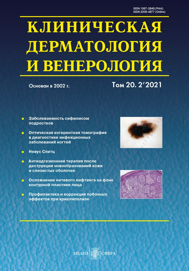
Опыт использования оптической когерентной томографии в комплексной диагностике инфекционных заболеваний ногтей
Журнал: Клиническая дерматология и венерология. 2021;20(2):44-49.
DOI: 10.17116/klinderma20212002144
Прочитано: 1648 раз
Резюме
Диагностика инфекционных заболеваний ногтей, в частности онихомикоза, нередко затруднена вследствие клинического сходства поражений инфекционной и неинфекционной природы, возможности их сочетания и большой вероятности ложноотрицательных результатов лабораторных исследований. Оптическая когерентная томография (ОКТ) — неинвазивный высокоразрешающий метод визуализации биотканей, приближающийся по информативности к гистологическому. Использование ОКТ позволяет оценивать компоненты ногтевого аппарата в норме и при некоторых патологических состояниях.
ЦЕЛЬ ИССЛЕДОВАНИЯ
Изучить возможность клинического использования ОКТ для диагностики некоторых инфекционных заболеваний ногтей.
МАТЕРИАЛ И МЕТОДЫ
Под наблюдением находились 47 пациентов с изолированным поражением ногтевых пластин стоп и/или кистей с подозрением на онихомикоз, у которых проведено обследование ногтевого аппарата, включавшее микроскопическое исследование с использованием 20% раствора калия гидроксида (КОН), культуральное исследование с использованием универсальной питательной среды Сабуро, исследование с использованием тест-системы Candiselect, а также ОКТ. При проведении ОКТ использовали визуализатор-топограф оптико-когерентный компьютеризированный для неинвазивного исследования внутренней структуры поверхностных тканей человека ВОК (ФГБНУ «Федеральный исследовательский центр Институт прикладной физики Российской академии наук», Россия).
РЕЗУЛЬТАТЫ
Томографические признаки онихомикоза выявлены у 35 пациентов, в том числе у 5 больных с отрицательными результатами исследования на дерматофиты. У 5 из них обнаружены ОКТ-признаки онихомикоза и псориаза ногтевых пластин. У 7 пациентов с подозрением на онихомикоз и отрицательными результатами исследования на дерматофиты при ОКТ-исследовании имелись признаки кандидозного поражения ногтей (при лабораторном обследовании диагноз подтвержден в 5 случаях). Противокандидозная терапия у этих больных оказала положительное действие, в том числе в случаях с отрицательными результатами лабораторного исследования. У 5 пациентов с отрицательными результатами исследований на дерматофиты и кандиды применение ОКТ позволило установить диагноз псевдомонадного поражения и провести адекватную терапию.
ЗАКЛЮЧЕНИЕ
Оптическая когерентная томография может эффективно использоваться в комплексной диагностике инфекционных заболеваний ногтей и оказывать существенную помощь в установлении диагноза при ложноотрицательных результатах лабораторных исследований на дерматофиты и кандиды, а также при сочетанной грибковой и псориатической патологии ногтевого аппарата.
Ключевые слова
- инфекционные заболевания ногтей
- диагностика
- оптическая когерентная томография
Дата поступления: 05.05.2020
Дата принятия в печать: 11.02.2021
Дата публикации: 15.04.2021
Введение
Инфекционные заболевания ногтей чрезвычайно распространены. Частота только онихомикозов составляет в популяции около 10% [1]. Этиологию заболеваний ногтевого аппарата, в том числе инфекционного происхождения, не всегда удается установить, что обусловлено неспецифичностью изменений ногтевых пластин при поражениях различной природы, возможностью сочетанной патологии, несовершенством лабораторной диагностики и невозможностью широкого применения морфологических исследований [2—4].
Технические сложности, возникающие при проведении биопсии в области ногтевого аппарата, высокий риск осложнений и остаточной деформации ограничивают ее использование практически только случаями подозрения на злокачественное новообразование и исключают многоочаговые и повторные исследования [3—5].
В качестве альтернативы биопсии при необходимости морфологического исследования ногтевого аппарата может рассматриваться оптическая когерентная томография (ОКТ) — высокоразрешающий (10—20 мк) неинвазивный метод визуализации структуры биотканей, приближающийся по информативности к методу эксцизионной биопсии. Этот метод основан на использовании в качестве зондирующего излучения низкоинтенсивного света ближнего инфракрасного диапазона и позволяет получать информацию о морфологическом состоянии биотканей на глубине до 1,5 мм в режиме реального времени. Структура ткани при ОКТ визуализируется за счет регистрации отраженной от внутритканевых элементов части зондирующего излучения [6].
Применение ОКТ позволяет визуализировать роговой и клеточные слои эпидермиса, сосочковый слой и верхнюю часть сетчатого слоя дермы с сосудами, кожные железы, компоненты ногтевого аппарата, а также качественно и количественно оценивать их структуру [7—10].
В силу специфических изменений оптических свойств структурных компонентов кожи при развитии в них определенных патоморфологических процессов ОКТ представляет возможность их верификации и на этом основании — использования метода в прижизненной диагностике некоторых дерматозов [11, 12].
Помимо отсутствия инвазивности, к достоинствам метода можно отнести компактность и доступную стоимость отечественных устройств, легкость в их эксплуатации, быстроту сканирования, возможность проведения повторных многократных исследований в разных зонах очага поражения и высокую информативность (информация, получаемая с помощью ОКТ, сопоставима с таковой при гистологическом исследовании). Волоконные системы доставки излучения отечественных томографов обеспечивают исследование любого участка кожи и ногтевого аппарата без длительной иммобилизации пациента, а результаты исследования практически не зависят от внешних факторов [8].
Возможности использования ОКТ в диагностике заболеваний ногтевого аппарата изучены недостаточно. В результате проведенных ранее исследований определены ОКТ-признаки компонентов здорового ногтевого аппарата (рис. 1, а), а также диагностические признаки поражения ногтей при псориазе, онихолизисе и онихомикозах [7, 9].

Рис. 1. ОКТ-изображения.
а — здоровая ногтевая пластина; б — ногтевая пластина с проявлениями онихомикоза: уменьшение интенсивности сигнала в пределах 1—4-го слоев изображения, характеризующих состояние собственно ногтевой пластины (1—3-й слои) и эпителия ногтевого ложа (4-й слой); линейные горизонтально и под углом ориентированные зоны сигнала высокой интенсивности в пределах 4—5-го слоев, являющиеся эквивалентом колоний гриба (отмечено стрелкой).
Цель исследования — изучить возможность клинического использования ОКТ для диагностики некоторых инфекционных заболеваний ногтей.
Материал и методы
Исследование проведено на базе Университетской клиники ФГБОУ ВО «Приволжский исследовательский медицинский университет» Минздрава России. В исследование включены 47 пациентов с изолированным поражением ногтевых пластин стоп и/или кистей с подозрением на онихомикоз.
Обследование больных включало микроскопическое исследование ногтевого аппарата с использованием 20% раствора гидроксида калия (КОН), культуральное исследование с использованием универсальной питательной среды Сабуро, исследование с применением тест-системы Candiselect, ОКТ.
При проведении ОКТ использовали визуализатор-топограф оптико-когерентный компьютеризированный для неинвазивного исследования внутренней структуры поверхностных тканей человека ВОК (ФГБНУ «Федеральный исследовательский центр Институт прикладной физики Российской академии наук», Россия, рег. уд. №ФС 022а2005/2035 от 05 августа 2005 г.) со следующими техническими характеристиками: длина волны излучения 920 нм, мощность 2 мВт, пространственное разрешение 15—20 мкм, глубина сканирования 1,5 мм, поперечный диапазон сканирования 1,8 мм, время получения двумерного изображения размером 200×200 точек 1,5 с. В качестве иммерсионного средства применяли гель для ультразвуковых исследований средней вязкости Медиагель.
Для работы с полученными изображениями использовали дополнительное программное обеспечение, разработанное в лаборатории биофотоники ИПФ РАН. Получаемые ОКТ-изображения представляют собой двумерные образы вертикального среза биоткани (ногтевого аппарата) в псевдоцветной коричневой палитре, имеющего ту же ориентацию, что и традиционный гистологический срез.
Визуально сравнивали ОКТ-изображения здоровых участков ногтевого аппарата, пограничной зоны и участков поражения с различной выраженностью проявлений. При тотальном поражении ногтевой пластины полученные изображения сравнивали с изображениями симметричной здоровой ногтевой пластины.
Оценку изображений ногтей осуществляли по следующим критериям: структурность, однородность, слоистость, контраст, характеристика границ слоев, интенсивность сигнала. Всего проанализировано 1209 ОКТ-изображений.
У каждого участника исследования получено письменное добровольное информированное согласие. Исследование одобрено этическим комитетом по проведению научных исследований ФГБОУ ВО НижГМА Минздрава России (протокол №2 от 18 февраля 2013 г.).
Результаты
В результате исследований у 35 пациентов с клиническим подозрением на грибковое поражение ногтей во всех полученных изображениях обнаружены ОКТ-признаки онихомикоза [7, 13, 14] (рис. 1, б), которые визуализировались в следующем виде:
— уменьшение интенсивности сигнала в пределах 1—4-го слоев изображения, характеризующих состояние собственно ногтевой пластины (1—3-й слои) и эпителия ногтевого ложа (4-й слой);
— увеличение суммарной высоты 2—4-го слоев (вследствие гиперкератоза);
— уменьшение сигнала в пределах 5-го слоя (эквивалент дермы в области ногтевого ложа);
— появление в пределах 4—5-го слоев линейных горизонтально и под углом ориентированных зон сигнала высокой интенсивности, являющихся эквивалентом колоний гриба.
При этом только в 30 случаях диагноз «онихомикоз» подтвержден микроскопическими и/или культуральными исследованиями, тогда как в остальных 5 случаях результаты повторных лабораторных исследований были отрицательными. Этим пациентам диагноз «онихомикоз» установлен только на основании клинических проявлений и данных ОКТ. Правомерность диагноза в дальнейшем подтверждена регрессом клинических проявлений поражения ногтей после проведения курса соответствующей противогрибковой терапии.
У 5 из 35 пациентов с клиническими и томографическими признаками онихомикоза при ОКТ-исследовании выявлены также признаки псориатического поражения ногтевого аппарата [7, 10] (рис. 2, а, б):
— увеличение суммарной высоты 2-го и 3-го слоев (проявление гиперкератоза);
— увеличение интенсивности сигнала и появление неоднородности в пределах 2-го и 3-го слоев (признак паракератоза);
— увеличение интенсивности сигнала в пределах 4-го и 5-го слоев с отсутствием их дифференцировки;
— увеличение высоты 5-го слоя с появлением в его пределах неоднородностей с регулярным чередованием вертикально расположенных зон с сигналом высокой и низкой интенсивности, характеризующихся выраженным контрастом между ними и формирующих специфический рисунок по типу «аркообразных сводов» (признак акантоза с папилломатозом).

Рис. 2. ОКТ-изображения.
а — ногтевая пластина с псориатическим поражением по типу масляного пятна: увеличение высоты 5-го слоя с регулярным чередованием вертикально расположенных зон с высокой и низкой интенсивностью сигнала, характеризующихся выраженным контрастом между ними и формирующих специфический рисунок по типу «аркообразных сводов» (признак акантоза с папилломатозом); б — участок ногтевой пластины с сочетанными признаками псориатического поражения и онихомикоза.
На основании выявленных изменений у больных нами диагностировано сочетанное поражение ногтевых пластин. При проведении комбинированного лечения (противогрибкового и противопсориатического) достигнут хороший терапевтический эффект. Необходимо отметить, что во всех 5 случаях псориатическое поражение ногтей имело изолированный характер, что, несомненно, затрудняло диагностику, а диагноз сопутствующего онихомикоза только у 2 пациентов подтвержден лабораторными исследованиями.
При обследовании 7 пациентов с клиническим подозрением на онихомикоз и отрицательными результатами лабораторного исследования на дерматофиты в 100% изображений обнаружены ОКТ-признаки кандидозного поражения ногтевого аппарата [9] (рис. 3, а):
— увеличение высоты 1-го слоя с неравномерным повышением интенсивности сигнала с визуализацией на этом фоне зон, характеризующихся сигналом низкой интенсивности;
— отсутствие визуализации 2—5-го слоев, что обусловлено значительным увеличением рассеивающих свойств поверхности ногтевой пластины вследствие ее шероховатой структуры;
— признаки выраженного воспаления в области заднего ногтевого валика и матрикса в виде уменьшения интенсивности сигнала в пределах 2—4-го слоев и отсутствия визуализации 5—8-го слоев (эквивалент корня ногтя и зоны матрикса) вследствие выраженного отека тканей (рис. 3, б).

Рис. 3. ОКТ-изображение ногтевого аппарата с проявлениями кандидозного поражения.
а — видимая часть ногтевой пластины в зоне поражения (отсутствие визуализации 2—5-го слоев, что обусловлено значительным увеличением рассеивающих свойств поверхности ногтевой пластины вследствие ее шероховатой структуры); б — задний ногтевой валик и матрикс (уменьшение интенсивности сигнала в пределах 2—4-го слоев и отсутствие визуализации 5—8-го слоев вследствие выраженного отека).
При этом только в 5 случаях диагноз удалось подтвердить культурально и микроскопически. Однако адекватная противокандидозная терапия дала позитивный эффект во всех случаях, в том числе у пациентов с ложноотрицательными результатами лабораторного исследования, диагноз кандидозного поражения которым установлен только на основании клинической картины и данных ОКТ.
У 5 из 47 пациентов с клиническим подозрением на грибковое поражение ногтей ОКТ не выявила томографических признаков онихомикоза или кандидоза. На всех полученных изображениях в пределах поражения визуализировалась щелевидная полость (щелевидная горизонтально ориентированная зона сигнала слабой интенсивности между 4-м и 5-м слоями изображения, ограниченная сверху линейной зоной сигнала высокой интенсивности) между собственно ногтевой пластиной и ногтевым ложем (рис. 4). Учитывая клинические особенности, а именно зеленоватый оттенок в окраске измененной части ногтевой пластины [13, 14], а также негативные результаты исследования на дерматофиты и кандиды, этим больным нами установлен диагноз псевдомонадного поражения. Проведенная соответствующая антибактериальная терапия дала положительный эффект.

Рис. 4. ОКТ-изображение ногтевой пластины с признаками псевдомонадной инфекции: щелевидная полость между собственно ногтевой пластиной и ногтевым ложем (щелевидная горизонтально ориентированная зона сигнала слабой интенсивности между 4-м и 5-м слоями изображения, ограниченная сверху линейной зоной сигнала высокой интенсивности).
Заключение
Результаты исследований свидетельствуют о том, что оптическая когерентная томография может эффективно использоваться в комплексной диагностике инфекционных заболеваний ногтей и оказать существенную помощь в установлении диагноза при ложноотрицательных результатах лабораторных исследований на дерматофиты и кандиды, а также при сочетанной грибковой и псориатической патологии ногтевого аппарата.
Участие авторов:
Концепция и дизайн исследования: Г.А. Петрова, М.С. Незнахина
Сбор и обработка материала: М.С. Незнахина, К.С. Петрова
Статистический анализ данных: М.С. Незнахина
Написание текста: Г.А. Петрова, М.С. Незнахина
Редактирование: Н.Н. Потекаев, Г.А. Петрова
Авторы заявляют об отсутствии конфликта интересов.
- Gupta AK, Drummond-Main C, Cooper EA, Brintnell W, Piraccini BM, Tosti A. Systematic review of nondermatophyte mold onychomycosis: diagnosis, clinical types, epidemiology, and treatment. Journal of the American Academy of Dermatology. 2012;66(3):494-502. https://doi.org/10.1016/j.jaad.2011.02.038
- Hay RJ, Baran R. Onychomycosis: a proposed revision of the clinical classification. Journal of the American Academy of Dermatology. 2011;65(6):1219-1227. https://doi.org/10.1016/j.jaad.2010.09.730
- Rothmund G, Sattler EC, Kaestle R, Fischer C, Haas CJ, Starz H, Welzel J. Confocal laser scanning microscopi as a new valuable tool in the diagnosis of onychomycosis — comparison of six diagnostic methods. Mycoses. 2013;56(1):47-55. https://doi.org/10.1111/j.1439-0507.2012.02198.x
- Radtke MA, Beikert FC, Augustin M. Nail psoriasis — a treatment challenge. Journal der Deutschen Dermatologischen Gesellschaft. 2013;11(3):203-219; quiz 220. https://doi.org/10.1111/ddg.12054
- Klaassen KM, Kerkhof PC, Pasch MC. Nail Psoriasis: a questionnaire-based survey. The British Journal of Dermatology. 2013;169(2):314-319. https://doi.org/10.1111/bjd.12354
- Welzel J. Optical coherence tomography. Venerologie, und Verwandte Gebiete. 2010;61(5):416-420. https://doi.org/10.1007/s00105-009-1879-x
- Петрова Г.А., Незнахина М.С., Гаранина О.Е., Шливко И.Л. Оценка эффективности бетаметазона с кальципотриолом при изолированном псориатическом поражении ногтевого аппарата методом оптической когерентной томографии. Вестник дерматологии и венерологии. 2013; 3:59-65.
- Петрова Г.А., Фирсова М.С., Шливко И.Л., Чекалкина О.Е., Зорькина М.В., Петрова К.С. Экспериментальная идентификация компонентов ОКТ-изображений ногтевого аппарата. Российский журнал кожных и венерических болезней. 2010;5:45-48.
- Sattler E, Kaestle R, Rothmund G, Welzel J. Confocal laser scanning microscopy, optical coherence tomography and transonychial water loss for in vivo investigation of nails. The British Journal of Dermatology. 2012;166(4): 740-746. https://doi.org/10.1111/j.1365-2133.2011.10730.x
- Aldahan AS, Chen LL, Fertig RM, Holmes J, Shah VV, Mlacker S, Hsu VM, Nouri K, Tosti A. Vascular Features of Nail Psoriasis Using Dynamic Optical Coherence Tomography. Skin Appendage Disorders. 2017;2(3-4):102-108. https://doi.org/10.1159/000449230
- Шливко И.Л., Петрова Г.А., Каменский В.А., Донченко Е.В., Зорькина М.В., Эллинский Д.О., Чекалкина О.Е. Комплексная оценка возрастных морфофункциональных особенностей кожи различной анатомической локализации. Российский журнал кожных и венерических болезней. 2011;6:51-55.
- Петрова Г.А., Дерпалюк Е.Н., Гладкова Н.Д., Никулин Н.К., Геликонов В.М., Геликонов Г.В., Иксанов Р.Р., Каменский В.А. Оптическая когерентная томография — эффективный метод прижизненного исследования структуры кожи в норме и при патологии. Экспериментальная и клиническая дерматокосметология. 2005;2:8-16.
- Piraccini BM, Balestri R, Starace M, Rech G. Nail digital dermoscopy (Onychoscopy) in the diagnosis of onychomycosis. Journal of the European Academy of Dermatology and Venereology: JEADV. 2013;27(4):509-513. https://doi.org/10.1111/j.1468-3083.2011.04323.x
- Brasch J, Shimanovich I. Persistent fingernail onychomycosis caused by Fusarium proliferatum in a healthy woman. Mycoses. 2012;55(1):86-89. https://doi.org/10.1111/j.1439-0507.2011.02041.x


Рекомендуем статьи по данной теме:
Возможности и ограничения искусственного интеллекта во флебологии: результаты систематического обзора литературы с метаанализом.Флебология. 2026;20(3):220-248
Совершенствование методов объективной диагностики острого риносинусита.Вестник оториноларингологии. 2026;91(4):88-91
Клинико-диагностические аспекты глаукоматозной кератопатии при врожденной глаукоме.Вестник офтальмологии. 2026;142(4):37-45
Современные возможности диагностики перипапиллярных неоваскулярных мембран. Сообщение 1. ОКТ-диагностические признаки.Вестник офтальмологии. 2026;142(4):26-36
Оптическая когерентная томография в диагностике эндотелиальной дистрофии роговицы Фукса.Вестник офтальмологии. 2026;142(4):5-13
Осведомленность врачей первичного звена Челябинской области о диагностике и лечении ревматоидного артрита.Профилактическая медицина. 2026;29(7):25-30
Эффективность внутрисосудистых методов исследования при лечении пациентов с «ложными» бифуркационными стенозами ствола левой коронарной артерии: результаты трехлетнего наблюдения.Кардиология и сердечно-сосудистая хирургия. 2026;19(4):32-39
Тактика хирурга при мукоцеле червеобразного отростка.Оперативная хирургия и клиническая анатомия (Пироговский научный журнал). 2026;10(2-2):103-108
Клинический случай диагностики и лечения экстраносоглоточной ангиофибромы.Вестник оториноларингологии. 2026;91(3):111-114
Инфекционный уретрит у мужчин: этиология, эпидемиология, диагностика и лечение (аналитический обзор).Клиническая дерматология и венерология. 2026;25(3):464-472