A critical case of postoperative duodenal fistula and retroperitoneal abscess after laparoscopic cholecystectomy: role of endoscopic fistula closure and multidisciplinary management
Abstract
Postoperative duodenal fistula is a rare but serious complication after cholecystectomy and nephrectomy. A case of a 58-year-old woman with chronic calculous cholecystitis and right-sided nephrolithiasis who developed a postoperative duodenal fistula with retroperitoneal abscess is presented. Initial conservative management and percutaneous drainage were performed. Endoscopic closure using TTS endoclips and an endoloop was performed after five months of persistent fistula. Complete fistula closure was achieved, confirmed by follow-up endoscopy and CT scan. Endoscopic closure with TTS endoclips and an endoloop is an effective, minimally invasive option for small chronic duodenal fistulas. Over-the-scope clips (OTSCs) were not used due to the small fistula size and well-formed granulation tissue, which allowed for successful closure with conventional endoscopic tools.
Keywords
- duodenal fistula
- retroperitoneal abscess
- laparoscopic cholecystectomy
- endoscopic closure
- TTS endoclips
- endoloop
Дата поступления: 08.05.2025
Дата принятия в печать: 03.09.2025
Дата публикации: 12.08.2026
Introduction
Duodenal perforation (DP) is considered a life-threatening injury. These injuries are reported in 4.3% of patients with abdominal trauma, with mortality rates ranging from 5.3% to 30% [1, 2]. DP is a type of polyetiologic lesion that occurs in patient with gastric and duodenal ulcer disease and is iatrogenic or posttraumatic in origin. CT with intravenous and oral contrast plays a key role in the diagnosis of DP [3, 4]. Treatment directly depends on the cause, duration, location, nature and general condition of the patient. Active conservative treatment under dynamic control is possible in closed or closed lesions and in clinically stable patients [5]. Patients with acute peritonitis, sepsis and intra-abdominal abscess require urgent surgical treatment. In recent decades, minimally invasive endoscopic treatment for DP has been considered an alternative to open surgical treatment [6—8] and is justified in practice.
Case description
A 58-year-old Caucasian woman (K.G., born in 1963) with a history of right-sided nephrolithiasis, terminal hydronephrosis and chronic calculous cholecystitis was initially treated at another medical center. She presented with severe right subcostal and epigastric pain radiating to the right scapula, accompanied by nausea, a bitter taste in the mouth and generalized weakness with elevated blood pressure (Table).
Timeline of diagnostic and therapeutic steps
Date | Event | Findings/Intervention | Outcome |
22.11.2021 | Ultrasound | Thickened gallbladder wall (2.5 mm), gallstones (7.0 mm, 2.6 mm) | Diagnosis: chronic calculous cholecystitis, right nephrolithiasis, terminal hydronephrosis |
24.11.2021 | Surgery | Laparoscopic cholecystectomy+right nephrectomy | — |
04.12.2021 | Post-op | Fever 38.6 °C, purulent drain output | Conservative treatment started |
13.12.2021 | CT scan | 26-mm retroperitoneal mass with air; 4-mm duodenal defect | Percutaneous drainage |
17.01.2022 | Labs/CT | Elevated CRP, leukocytes; 22-mm retroperitoneal fluid | Second percutaneous drainage |
23.03.2022 | ICU admission | Duodenal perforation, retroperitoneal abscess, sepsis | Intensive care |
09.04.2022 | Endoscopy | 0.4×0.6 cm duodenal fistula | TTS endoclips+endoloop closure |
01.05.2022 | Follow-up CT | No contrast extravasation; fibrotic tissue | Complete fistula closure confirmed |
The initial (22.11.2021) diagnostic ultrasound revealed a thickened gallbladder wall (2.5 mm) and multiple gallstones (7.0 mm and 2.6 mm). A diagnosis of chronic calculous cholecystitis, right-sided nephrolithiasis and terminal hydronephrosis was made.
After a short preoperative preparation (23.11.2021), a «laparoscopic cholecystectomy» (LCE) and a one-stage «laparoscopic right-sided nephrectomy» were performed the next day under multicomponent intravenous anaesthesia with endotracheal intubation.
On postoperative day 10, the patient developed a fever of 38.6 °C, along with general weakness and malaise, and purulent discharge was observed from the intraperitoneal drain. She received prolonged conservative management, including antibiotic therapy and supportive care.
A CT scan performed on 13.12.2021 revealed a 26-mm mass with air bubbles in the right retroperitoneal region, along with edema and infiltration of the surrounding adipose tissue. A 4-mm defect was identified in the right posterolateral wall of the second duodenal segment, with contrast extravasation into the retroperitoneal mass. Thickening of the adjacent peritoneal layers was also noted (Fig. 1). The patient underwent percutaneous drainage of the right retroperitoneal space and was discharged for outpatient care.
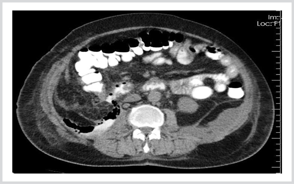
Fig. 1. Extravasation of orally administered contrast medium from the duodenal defect into a mass within the right retroperitoneal region was observed.
Laboratory results on 17.01.2022 showed elevated CRP (141.6 mg/L), creatinine (103 µmol/L), ESR (105 mm/h), platelets (520·109/L), leukocytes (13.0·109/L), and decreased hemoglobin (6.7 g/dL). A repeat CT scan on the same day revealed a well-demarcated fluid mass (22 mm) with air bubbles in the right retroperitoneal region, significant peritoneal thickening, and surrounding adipose tissue edema. The patient underwent a second percutaneous drainage procedure on 18.01.2022 and continued outpatient care.
On 23.03.2022, the patient was admitted to the intensive care unit with postoperative duodenal perforation, functional fistula, retroperitoneal abscess, and sepsis. Laboratory results at admission included CRP 129.6 mg/L, albumin 3.56 g/dL, hemoglobin 8.0 g/dL, platelet count 584·109/L, and ESR 91 mm/h.
A CT scan on 28.03.2022 revealed a 45-mm retroperitoneal abscess extending from the subdiaphragmatic area to the iliac region and descending along the lumbar muscle, with air bubbles present. Oral contrast extravasation into the retroperitoneal space was noted on the right. Additional focal abscesses with air bubbles were identified on the anterior and lateral bladder walls in the left iliac region and pelvis, with the drainage tube tip visible anterior to the bladder (Fig. 2).
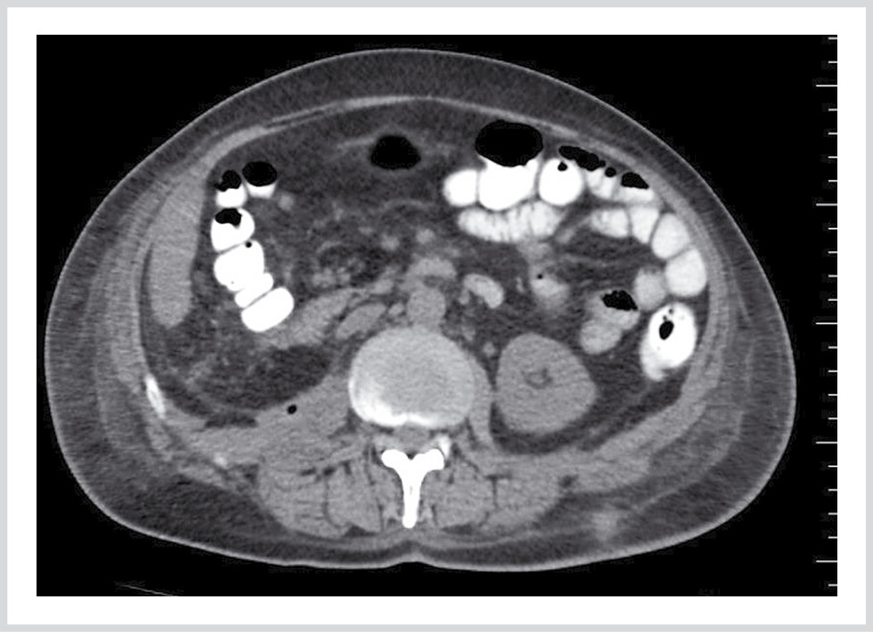
Fig. 2. Extravasation of the oral contrast agent into the retroperitoneal space was observed.
Despite five months of conservative treatment, including two percutaneous drainages, no attempt had been made to close the fistula prior to presentation at our center on 09.04.2022. Endoscopic examination revealed a 0.4×0.6 cm fistula surrounded by granulation tissue on the posterior wall of the second duodenal segment (Fig. 3). TTS endoclips (Olympus, Japan) were applied along the edges of the fistula (Fig. 4, a), and compression with an endoloop (Olympus, Japan) successfully achieved fistula closure (Fig. 4, b, c). One month later, repeat endoscopy confirmed complete fistula closure with scar formation (Fig. 5). A follow-up CT scan with oral contrast on 01.05.2022 showed no contrast extravasation or retroperitoneal leakage, although fibrotic tissue was observed around the duodenum (Fig. 6).
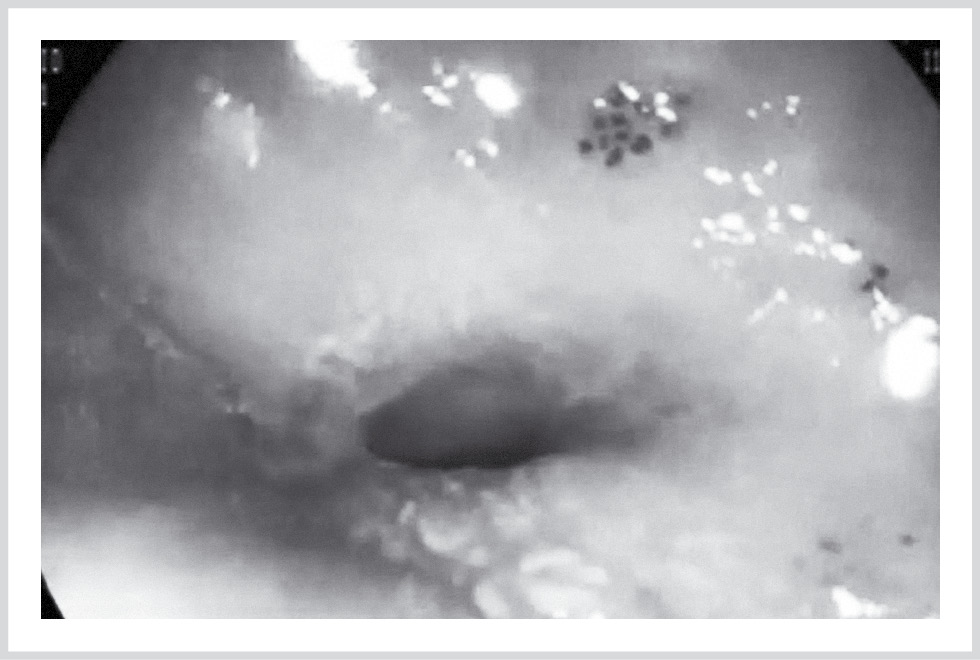
Fig. 3. A fistula tunnel was identified in the posterior wall of the duodenum.
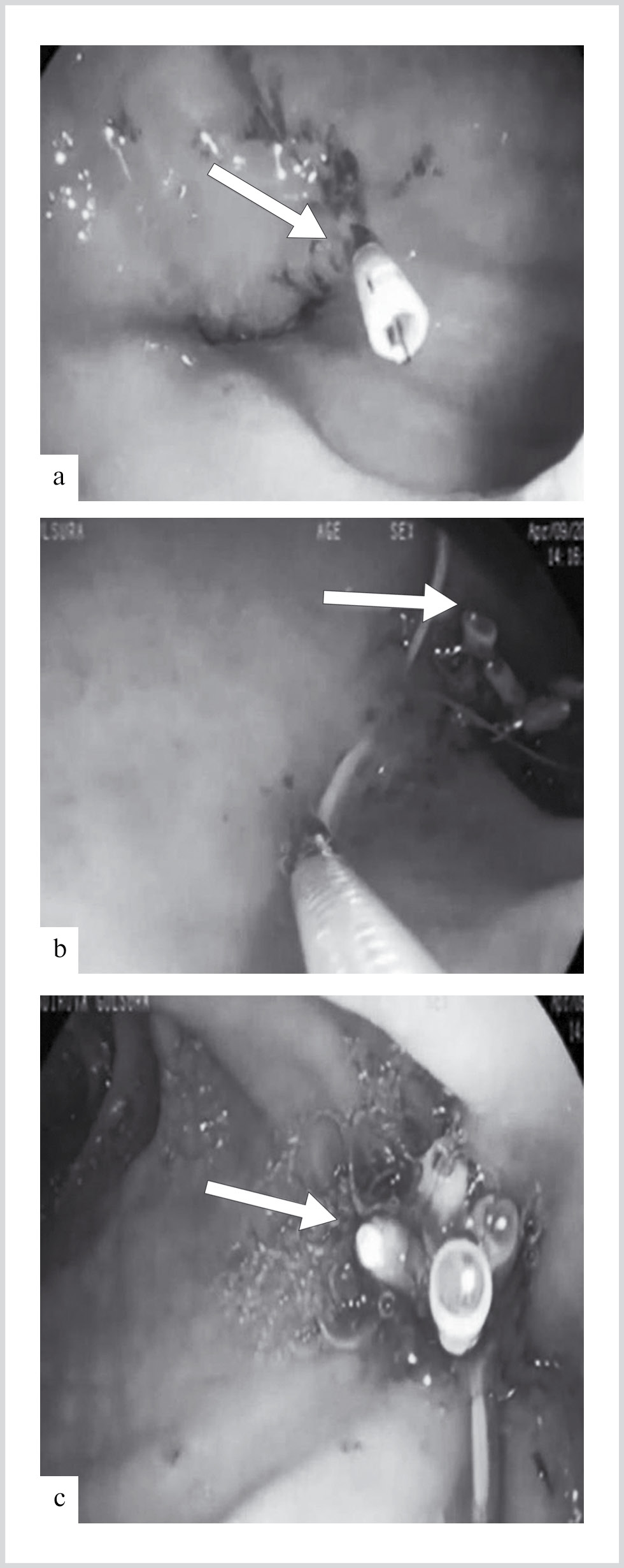
Fig. 4. Stages of endoscopic closure using TTS clips applied to the edges of the fistula (a—c).
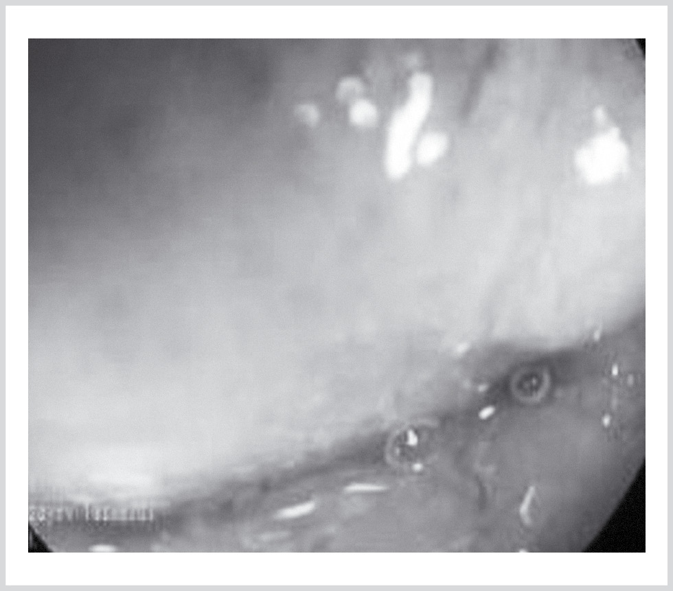
Fig. 5. Scar tissue at the site of the opening of the fistula on the posterior wall of the duodenum.
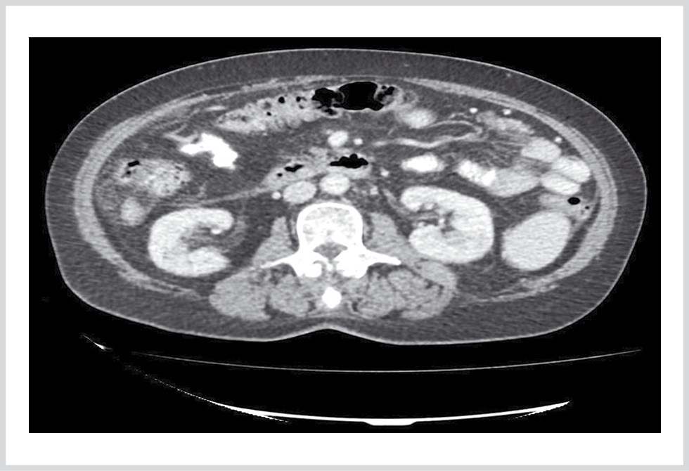
Fig. 6. Contrast-enhanced CT of the abdomen showing fibrous thickening extending from the second segment of the duodenum to Gerota’s fascia in the retroperitoneal region, with no extravasation of oral contrast observed.
Discussion
Duodenal perforation (DP) can result from various causes, including duodenal diverticulum, ischemia, infectious and autoimmune diseases (such as Crohn’s disease, scleroderma, and vasculitis), malignancies, chemotherapy, and gallstone disease [1, 2]. Iatrogenic DP typically occurs due to improper surgical techniques, particularly during tissue dissection [3, 4]. This complication is most commonly observed after laparoscopic procedures, with an incidence of 0.015% [5, 6]. Coagulation necrosis of the duodenal wall may occur due to excessive cauterization, and perforation is often not detected intraoperatively. Symptoms typically manifest several days postsurgery [7, 8].
Treatment for DP includes conservative management, endoscopic intervention, and surgery [9—11]. Concurrently, intensive resuscitation, infection control, and supportive care are essential. While surgical repair can definitively close the perforation, its invasiveness and associated risks are significant drawbacks. In the present case, perforation symptoms emerged 10 days postsurgery, and an endoscopic approach was employed to close the fistula [12, 13].
Endoscopic treatment is a minimally invasive and effective method, particularly when performed within the first 24 hours, before significant inflammatory changes occur in the surrounding tissue [14, 15]. Through-the-scope (TTS) endoclips are highly effective for small duodenal perforations (≤1 cm) [16, 17]. For larger perforations, over-the-scope clips (OTSCs) can grasp more tissue and close defects by compressing all the tissue layers. An OTSC is typically used for perforations between 1-and 3 cm in diameter [18, 19].
In our case, owing to the delayed presentation and the surrounding scar tissue, a TTS clip alone was insufficient for complete closure. Therefore, after placing the endoclips were placed, an endoloop was used to secure them and eliminate any gap between the clips, ensuring reliable and permanent closure. This combined approach was successful in completely sealing the perforation. The use of both endoclips and endoloops allowed for complete closure of the fistula, demonstrating the effectiveness of this technique, which would not have been achieved with endoclips alone.
Funding
This research received no external funding.
Consent for Publication:
The patient provided written informed consent for the publication of any potentially identifiable images included in this article.
Participation of authors:
Concept and design of the study — Rzayev T., Khidirova N.,
Data collection and processing — Rzayev T., Khidirova N., Ibrahimova A., Bayramov N.
Text writing — Rzayev T., Khidirova N., Ibrahimova A.
Editing — Ibrahimova A., Bayramov N.
- García Santos E, Soto Sánchez A, Verde JM, Marini CP, Asensio JA, Petrone P. Duodenal injuries due to trauma: Review of the literature. Cir Esp. 2015;93(2):68-74. https://doi.org/10.1016/j.ciresp.2014.08.004
- Asensio JA, Petrone P, Roldán G, Pak-art R, Salim A. Pancreatic and duodenal injuries: complex and lethal. Scand J Surg. 2002;91:81-86. https://doi.org/10.1177/145749690209100113
- Mendoza J, Tovar G, Álvarez R. Hepatoduodenal fistula closure diagnosed and characterized Ecoendoscopically (EUS) and managed by OTSC CLIP OVESCO: A case report. Int J Surg Case Rep. 2021;84:106093. https://doi.org/10.1016/j.ijscr.2021.106093
- Musicant ME, Thompson JC. The emergency management of lateral duodenal fistula by pancreaticoduodenectomy. Surg Gynecol Obstet. 1969;128:108-114.
- Charabaty-Pishvaian A, Al-Kawas F. Endoscopic treatment of duodenal perforation using a clipping device. Southern Med J. 2004;97:190-193. https://doi.org/10.1097/01.SMJ.0000091031.77846.B6
- Bianchi A, Solduga C, Ubach M. Percutaneous obliteration of a chronic duodenal fistula. Br J Surg. 1988;75:572. https://doi.org/10.1002/bjs.1800750624
- Cotton PB, Lehman G, Vennes J, Geenen JE, Russell RC, Meyers WC, Liguory C, Nickl N. Endoscopic sphincterotomy complications and their management: an attempt at consensus. Gastrointest Endosc. 1991;37:383-393. https://doi.org/10.1016/s0016-5107(91)70740-2
- Gaillard M, Dupond-Athenor A, Donatelli G, I Dagher. Conservative endoscopic management of a large duodenal defect after cholecystectomy. J Visc Surg. 2017;154:379-381. https://doi.org/10.1016/j.jviscsurg.2017.06.009
- Gregorios A Paspatis, Marianna Arvanitakis, Jean-Marc Dumonceau, Marc Barthet, Brian Saunders, Stine Ydegaard Turino, Angad Dhillon, Maria Fragaki, Jean-Michel Gonzalez, Alessandro Repici, Roy LJ van Wanrooij, Jeanin E van Hooft. Diagnosis and management of iatrogenic endoscopic perforations: European Society of Gastrointestinal Endoscopy (ESGE) Position Statement. Endoscopy. 2014;46:693-711. https://doi.org/10.1055/a-1222-3191
- Habr-Gama A, Waye JD. Complications and hazards of gastrointestinal endoscopy. World J Surg. 1989;13:193-201. https://doi.org/10.1007/BF01658399
- Haider S, Kahaleh M. The use of endoscopic clipping devices in the treatment of iatrogenic duodenal perforation. Gastroenterol Hepatol (NY). 2010;6:660-661.
- Isaguirre J, Gutiérrez S, Ongay R, Ravier RP. Endoscopic treatment of duodenal perforation following laparoscopic cholecystectomy. Endoscopy. 2008;40(Suppl 2):E138. https://doi.org/10.1055/s-2007-995731
- Kaneko T, Akamatsu T, Shimodaira K, Ueno T, Gotoh A, Mukawa K, Nakamura N, Kiyosawa K. Nonsurgical treatment of duodenal perforation by endoscopic repair using a clipping device. Gastrointest Endosc. 1999;50:410-414. https://doi.org/10.1053/ge.1999.v50.97235
- Katsinelos P, Paroutoglou G, Papaziogas B. Treatment of a duodenal perforation secondary to an endoscopic sphincterotomy with clips. World J Gastroenterol. 2005;11:6232-6234. https://doi.org/10.3748/wjg.v11.i39.6232
- Machado NO. Management of duodenal perforation post-endoscopic retrograde cholangiopancreatography: When and whom to operate and what factors determine the outcome? JOP. 2012;13:18-25.
- Mangiavillano B, Viaggi P, Masci E. Endoscopic closure of acute iatrogenic perforations during diagnostic and therapeutic endoscopy in the gastrointestinal tract using metallic clips: a literature review. J Dig Dis. 2010;11(1):12-18. https://doi.org/10.1111/j.1751-2980.2009.00414.x
- Melita G, Currò G, Iapichino G, Princiotta S, Cucinotta E. Duodenal perforation secondary to biliary stent dislocation: a case report and review of the literature. Chir Ital. 2005;57(3):385-388.
- Sabharwal T, Hatzidakis A, Papageorgiou G, Dourado R, Adam A. Trans-oral embolization of a postoperative duodenocutaneous fistula. Clin Radiol. 2004;59:102-104. https://doi.org/10.1016/j.crad.2003.08.004
- von Renteln D, Rudolph HU, Schmidt A, Vassiliou MC, Caca K. Endoscopic closure of duodenal perforations by using an over-the-scope clip: a randomized, controlled porcine study. Gastrointest Endosc. 2010;71:131-138. https://doi.org/10.1016/j.gie.2009.07.006
Recommended articles:
Abdominal Lymphatic System Response on Endosurgical Pneumoperitoneum.Journal of Venous Disorders. 2026;20(3):200-210
Clinical efficacy of combined subhepatic space drainage during laparoscopic cholecystectomy in patients with gangrenous cholecystitis.Endoscopic Surgery. 2026;32(4):46-53
Remaxol in perioperative infusion therapy for patients with calculous obstructive jaundice: a multiple-center observational study.Pirogov Russian Journal of Surgery. 2026;(6):22-28
Non-pharmacological pain relief after laparoscopic cholecystectomy: the role of pulmonary recruitment maneuver.Endoscopic Surgery. 2026;32(2):41-41
Analysis of the outcomes of standardized laparoscopic cholecystectomy.Endoscopic Surgery. 2026;32(2):28-34
Laparoscopic subtotal cholecystectomy for severe cholecystitis.Pirogov Russian Journal of Surgery. 2025;(7):67-73
Standardized laparoscopic cholecystectomy: methodology for the formation of intraoperative safety algorithms.Endoscopic Surgery. 2025;31(1):25-30
Transversus abdominis plane block for laparoscopic cholecystectomy in short-term acute care hospital.Pirogov Russian Journal of Surgery. 2024;(12):13-19