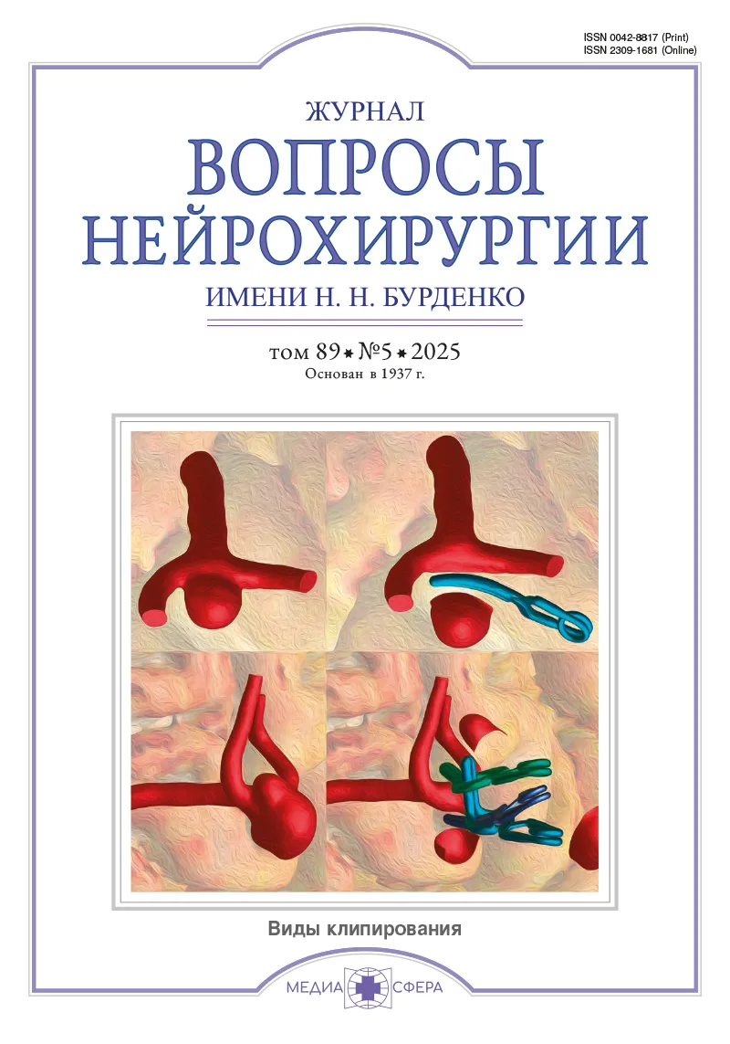
Хирургическое лечение внутримозговых опухолей лобной доли. Современное состояние проблемы. Часть I: двигательная функциональная система
Журнал: Вопросы нейрохирургии имени Н.Н. Бурденко. 2025;89(5):115-121.
DOI: 10.17116/neiro202589051115
Прочитано: 1664 раза
Резюме
Наиболее часто в обыденной клинической практике нейрохирург имеет дело с глиальными опухолями, локализованными именно в лобной доле. Существует распространенное в широких кругах мнение, что глиому лобной доли может удалить практически любой нейрохирург, независимо от опыта и уровня микрохирургических навыков. Однако, работая в федеральном центре, приходится наблюдать, как сотрудникам нередко приходится реоперировать пациентов с глиомами лобной доли, которым была выполнена операция в других лечебных учреждениях. Поэтому, на наш взгляд, лечение пациентов с данной патологией является не такой простой, как кажется на первый взгляд, проблемой нейрохирургии глиальных опухолей, а частота послеоперационных осложнений у данной категории больных остается высокой. Целью нашей работы был анализ литературы, касающейся как анатомо-функциональных аспектов хирургии глиом лобной доли, так и практических нюансов их хирургического лечения. Результаты анализа показали, что двигательная функциональная система лобной доли представлена несколькими корковыми зонами, с помощью ассоциативных волокон они объединяются в сложно организованную сеть функциональных связей, повреждение которых может приводить к послеоперационному двигательному дефициту.
Ключевые слова
- лобная доля мозга
- внутримозговые глиальные опухоли
- нейроонкология
Дата поступления: 11.03.2025
Дата принятия в печать: 05.06.2025
Дата публикации: 16.10.2025
- Banerjee K, Núñez FJ, Haase S, McClellan BL, Faisal SM, Carney SV, Yu J, Alghamri MS, Asad AS, Candia AJN, Varela ML, Candolfi M, Lowenstein PR, Castro MG. Current Approaches for Glioma Gene Therapy and Virotherapy. Frontiers in Molecular Neuroscience. 2021;14:621831. https://doi.org/10.3389/fnmol.2021.621831
- Larjavaara S, Mäntylä R, Salminen T, Haapasalo H, Raitanen J, Jääskeläinen J, Auvinen A. Incidence of gliomas by anatomic location. Neuro-Oncology. 2007;9(3):319-325. https://doi.org/10.1215/15228517-2007-016
- Low JT, Ostrom QT, Cioffi G, Neff C, Waite KA, Kruchko C, Barnholtz-Sloan JS. Primary brain and other central nervous system tumors in the United States (2014-2018): A summary of the CBTRUS statistical report for clinicians. Neuro-Oncology Practice. 2022;9(3):165-182. https://doi.org/10.1093/nop/npac015
- Ostrom QT, Patil N, Cioffi G, Waite K, Kruchko C, Barnholtz-Sloan JS. CBTRUS Statistical Report: Primary Brain and Other Central Nervous System Tumors Diagnosed in the United States in 2013-2017. Neuro-Oncology. 2020;22(12 Suppl 2):iv1-iv96. https://doi.org/10.1093/neuonc/noaa200
- Ostrom QT, Price M, Neff C, Cioffi G, Waite KA, Kruchko C, Barnholtz-Sloan JS. CBTRUS Statistical Report: Primary Brain and Other Central Nervous System Tumors Diagnosed in the United States in 2015-2019. Neuro-Oncology. 2022;24(5):v1-v95. https://doi.org/10.1093/neuonc/noac202
- Dum RP, Strick PL. Motor areas in the frontal lobe of the primate. Physiology & Behavior. 2002;77(4-5):677-682. https://doi.org/10.1016/s0031-9384(02)00929-0
- Rizzolatti G, Fogassi L, Gallese V. Motor and cognitive functions of the ventral premotor cortex. Current Opinion in Neurobiology. 2002;12(2):149-154. https://doi.org/10.1016/s0959-4388(02)00308-2
- Sughrue ME. The Glioma Book. 2019th ed. Thieme Verlag; 2019. https://doi.org/10.1055/b-006-160177
- Laplane D, Talairach J, Meininger V, Bancaud J, Orgogozo JM. Clinical consequences of corticectomies involving the supplementary motor area in man. Journal of the Neurological Sciences. 1977;34(3):301-314. https://doi.org/10.1016/0022-510x(77)90148-4
- Rossi M, Fornia L, Puglisi G, Leonetti A, Zuccon G, Fava E, Milani D, Casarotti A, Riva M, Pessina F, Cerri G, Bello L. Assessment of the praxis circuit in glioma surgery to reduce the incidence of postoperative and long-term apraxia: a new intraoperative test. Journal of Neurosurgery. 2019;130(1):17-27. https://doi.org/10.3171/2017.7.JNS17357
- Cerri G, Shimazu H, Maier MA, Lemon RN. Facilitation from ventral premotor cortex of primary motor cortex outputs to macaque hand muscles. Journal of Neurophysiology. 2003;90(2):832-842. https://doi.org/10.1152/jn.01026.2002
- Fornia L, Rossi M, Rabuffetti M, Leonetti A, Puglisi G, Viganò L, Simone L, Howells H, Bellacicca A, Bello L, Cerri G. Direct Electrical Stimulation of Premotor Areas: Different Effects on Hand Muscle Activity during Object Manipulation. Cerebral Cortex. 2020;30(1):391-405. https://doi.org/10.1093/cercor/bhz139
- Johnson PB, Ferraina S, Bianchi L, Caminiti R. Cortical networks for visual reaching: physiological and anatomical organization of frontal and parietal lobe arm regions. Cerebral Cortex. 1996;6(2):102-119. https://doi.org/10.1093/cercor/6.2.102
- Pesaran B, Nelson MJ, Andersen RA. Dorsal premotor neurons encode the relative position of the hand, eye, and goal during reach planning. Neuron. 2006;51(1):125-134. https://doi.org/10.1016/j.neuron.2006.05.025
- Vingerhoets G, Honoré P, Vandekerckhove E, Nys J, Vandemaele P, Achten E. Multifocal intraparietal activation during discrimination of action intention in observed tool grasping. Neuroscience. 2010;169(3):1158-1167. https://doi.org/10.1016/j.neuroscience.2010.05.080
- Gerloff C, Corwell B, Chen R, Hallett M, Cohen LG. Stimulation over the human supplementary motor area interferes with the organization of future elements in complex motor sequences. Brain. 1997;120(Pt 9):1587-1602. https://doi.org/10.1093/brain/120.9.1587
- Brinkman C. Supplementary motor area of the monkey’s cerebral cortex: short- and long-term deficits after unilateral ablation and the effects of subsequent callosal section. Journal of Neuroscience. 1984;4(4):918-929. https://doi.org/10.1523/JNEUROSCI.04-04-00918.1984
- Kim YH, Kim CH, Kim JS, Lee SK, Han JH, Kim CY, Chung CK. Risk factor analysis of the development of new neurological deficits following supplementary motor area resection. Journal of Neurosurgery. 2013;119(1):7-14. https://doi.org/10.3171/2013.3.JNS121492
- Krainik A, Lehéricy S, Duffau H, Vlaicu M, Poupon F, Capelle L, Cornu P, Clemenceau S, Sahel M, Valery CA, Boch AL, Mangin JF, Bihan DL, Marsault C. Role of the supplementary motor area in motor deficit following medial frontal lobe surgery. Neurology. 2001;57(5):871-878. https://doi.org/10.1212/wnl.57.5.871
- Krainik A, Lehéricy S, Duffau H, Capelle L, Chainay H, Cornu P, Cohen L, Boch AL, Mangin JF, Le Bihan D, Marsault C. Postoperative speech disorder after medial frontal surgery: role of the supplementary motor area. Neurology. 2003;60(4):587-594. https://doi.org/10.1212/01.wnl.0000048206.07837.59
- Russell SM, Kelly PJ. Incidence and clinical evolution of postoperative deficits after volumetric stereotactic resection of glial neoplasms involving the supplementary motor area. Neurosurgery. 2003;52(3):506-516. https://doi.org/10.1227/01.neu.0000047670.56996.53
- Vassal M, Charroud C, Deverdun J, Le Bars E, Molino F, Bonnetblanc F, Boyer A, Dutta A, Herbet G, Moritz-Gasser S, Bonafé A, Duffau H, de Champfleur NM. Recovery of functional connectivity of the sensorimotor network after surgery for diffuse low-grade gliomas involving the supplementary motor area. Journal of Neurosurgery. 2017;126(4):1181-1190. https://doi.org/10.3171/2016.4.JNS152484
- Davidoff RA. The pyramidal tract. Neurology. 1990;40(2):332-339. https://doi.org/10.1212/wnl.40.2.332
- Ebeling U, Reulen HJ. Subcortical topography and proportions of the pyramidal tract. Acta Neurochirurgica. 1992;118(3-4):164-171. https://doi.org/10.1007/BF01401303
- Corbetta M, Shulman GL. Control of goal-directed and stimulus-driven attention in the brain. Nature Reviews Neuroscience. 2002;3(3):201-215. https://doi.org/10.1038/nrn755
- Halsband U, Passingham R. The role of premotor and parietal cortex in the direction of action. Brain Research. 1982;240(2):368-372. https://doi.org/10.1016/0006-8993(82)90239-6
- Leiguarda RC, Marsden CD. Limb apraxias: higher-order disorders of sensorimotor integration. Brain. 2000;123(5):860-879. https://doi.org/10.1093/brain/123.5.860
- Быканов А.Е., Пицхелаури Д.И., Баталов А.И., Пронин И.Н., Шкарубо М.А., Добровольский Г.Ф., Кобяков Г.Л., Буклина С.Б., Пучков В.Л., Захарова Н.Е., Смирнов А.С., Саникидзе А.З., Гольбин Д.А., Погосбекян Э.Л., Кудиева Э.С., Шкатова А.М., Потапов А.А. Хирургическая анатомия периинсулярных ассоциативных проводящих путей. Часть I. Система верхнего продольного пучка. Вопросы нейрохирургии им. Н.Н. Бурденко. 2017;81(1):26-38. https://doi.org/10.17116/neiro201780726-38
- De Pisapia N, Sandrini M, Braver TS, Cattaneo L. Integration in Working Memory: A Magnetic Stimulation Study on the Role of Left Anterior Prefrontal Cortex. PLoS ONE. 2012;7(8):e43731. https://doi.org/10.1371/journal.pone.0043731
- Knapen T, Brascamp J, Pearson J, van Ee R, Blake R. The role of frontal and parietal brain areas in bistable perception. Journal of Neuroscience. 2011;31(28):10293-10301. https://doi.org/10.1523/JNEUROSCI.1727-11.2011
- Catani M, Thiebaut de Schotten M. Atlas of Human Brain Connections. OUP Oxford; 2012.
- Kinoshita M, de Champfleur NM, Deverdun J, Moritz-Gasser S, Herbet G, Duffau H. Role of fronto-striatal tract and frontal aslant tract in movement and speech: an axonal mapping study. Brain Structure & Function. 2015;220(6):3399-3412. https://doi.org/10.1007/s00429-014-0863-0
- Rojkova K, Volle E, Urbanski M, Humbert F, Dell’Acqua F, Thiebaut de Schotten M. Atlasing the frontal lobe connections and their variability due to age and education: a spherical deconvolution tractography study. Brain Structure & Function. 2016;221(3):1751-1766. https://doi.org/10.1007/s00429-015-1001-3
- Dick AS, Garic D, Graziano P, Tremblay P. The frontal aslant tract (FAT) and its role in speech, language and executive function. Cortex. 2019;111: 148-163. https://doi.org/10.1016/j.cortex.2018.10.015
- La Corte E, Eldahaby D, Greco E, Aquino D, Bertolini G, Levi V, Ottenhausen M, Demichelis G, Romito LM, Acerbi F, Broggi M, Schiariti MP, Ferroli P, Bruzzone MG, Serrao G. The Frontal Aslant Tract: A Systematic Review for Neurosurgical Applications. Frontiers in Neurology. 2021;12:641586. https://doi.org/10.3389/fneur.2021.641586


Рекомендуем статьи по данной теме:
Хирургическое лечение внутримозговых опухолей лобной доли. Современное состояние проблемы. Часть III: клинические, нейрофизиологические и хирургические нюансы.Вопросы нейрохирургии имени Н.Н. Бурденко. 2026;90(1):108-115
Конъюгированные молекулы на основе аптамеров в экспериментальных и клинических подходах к терапии глиобластомы.Вопросы нейрохирургии имени Н.Н. Бурденко. 2025;89(6-2):44-49
Пилоцитарные астроцитомы с агрессивным клиническим течением. Серия пациентов ФГАУ «НМИЦ нейрохирургии им. акад. Н.Н. Бурденко» Минздрава России.Вопросы нейрохирургии имени Н.Н. Бурденко. 2025;89(6-2):29-37
Хирургическое лечение внутримозговых опухолей лобной доли. Часть II: речевая функциональная система.Вопросы нейрохирургии имени Н.Н. Бурденко. 2025;89(6):114-119