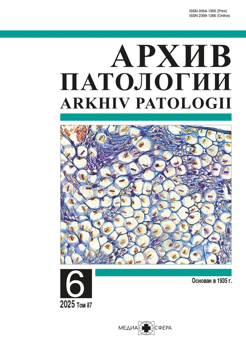
Васкуляризация эндометрия при гиперплазии без атипии
Резюме
Различные доброкачественные изменения эндометрия отличаются особенностями васкуляризации. Развитие рака эндометрия также сопровождается изменениями микроциркуляторного русла. Выявление васкуляризации гиперплазированного эндометрия без признаков атипии, выраженной значительно больше, чем описано в литературе, послужило основанием для этой работы.
ЦЕЛЬ ИССЛЕДОВАНИЯ
Оценить состояние сосудистого русла эндометрия при простой гиперплазии без атипии и сравнить полученные результаты с данными литературы.
МАТЕРИАЛ И МЕТОДЫ
Методом световой микроскопии с применением иммуногистохимической реакции с антителами к антигену CD34 и морфометрии у 31 женщины изучена васкуляризация эндометрия при его простой гиперплазии без атипии.
РЕЗУЛЬТАТЫ
Выявлена разная степень выраженности, но всегда значительная васкуляризация эндометрия при его простой гиперплазии без атипии. Сосуды занимали 5,97±4,18% от площади среза, при этом их количество на 1 мм2 было равно 391±180. Эти данные намного превышают большинство представленных в литературе результатов других исследований, которые отличаются в несколько раз даже между собой.
ЗАКЛЮЧЕНИЕ
Эндометрий при простой гиперплазии без атипии характеризуется различным, часто очень высоким уровнем васкуляризации, реальные средние показатели намного превосходят таковые, указанные в большинстве источников литературы, которые также очень противоречивы. Необходимо критически относиться к результатам, указывающим на особенности ангиогенеза и васкуляризации эндометрия при его гиперплазии.
Ключевые слова
- гиперплазия эндометрия
- васкуляризация эндометрия
- ангиогенез
Дата поступления: 13.05.2025
Дата принятия в печать: 25.06.2025
Дата публикации: 02.12.2025
- Mori H, Nishida H, Kusaba T, et al. Clinicopathological correlations of endometrioid and clear cell carcinomas in the uterus and ovary. Medicine (Baltimore). 2023;102(37):e35301. https://doi.org/10.1097/MD.0000000000035301
- Wang XX, Hua T, Wang HB. Estrogen receptor-related receptor γ uppresses hypoxia-induced angiogenesis by regulating VEGFA in endometrial cancer. Gynecol Endocrinol. 2023;39(1):2264411. https://doi.org/10.1080/09513590.2023.2264411
- Yoshino O, Ono Y. The physiological role of macrophages in reproductive organs. Reprod Med Biol. 2025;24(1):e12637. https://doi.org/10.1002/rmb2.12637
- Yetkin-Arik B, Kastelein AW, Klaassen I, et al. Angiogenesis in gynecological cancers and the options for anti-angiogenesis therapy. Biochim Biophys Acta Rev Cancer. 2021;1875(1):188446. https://doi.org/10.1016/j.bbcan.2020.188446
- Майбородин И.В., Гончаров М.А., Шевела А.И. и др. Ангиогенез при раке эндометрия: клиническое и биологическое значение. Сибирский онкологический журнал. 2024;23(4):172-185. https://doi.org/10.21294/1814-4861-2024-23-4-172-185
- Ozalp S, Yalcin OT, Acikalin M, et al. Microvessel density (MVD) as a prognosticator in endometrial carcinoma. Eur J Gynaecol Oncol. 2003;24(3-4):305-308.
- Guşet G, Costi S, Lazăr E, et al. Expression of vascular endothelial growth factor (VEGF) and assessment of microvascular density with CD34 as prognostic markers for endometrial carcinoma. Rom J Morphol Embryol. 2010;51(4):677-682.
- Kilinç E, Bahar AY. The Value of Intratumoral and Extratumoral Microvessel Density for the Tumor-dominated Area in the Endometrial Carcinoma. Appl Immunohistochem Mol Morphol. 2022;30(7):501-508. https://doi.org/10.1097/PAI.0000000000001044
- Drocaş I, Crăiţoiu Ş, Stepan AE, et al. The analysis of hormonal status and vascular and cell proliferation in endometrioid endometrial adenocarcinomas. Rom J Morphol Embryol. 2022;63(1):113-120. https://doi.org/10.47162/RJME.63.1.11
- Kaku T, Kamura T, Kinukawa N, et al. Angiogenesis in endometrial carcinoma. Cancer. 1997;80(4):741-747. https://doi.org/10.1002/(sici)1097-0142(19970815)80:4<741::aid-cncr13>3.0.co;2-t
- Bhosale NM, Arakeri SU, Reddy AK, Mudanur SR. Endometrial blood vessel morphometry in patients presenting with abnormal uterine bleeding. Indian J Pathol Microbiol. 2022;65(4):844-850. https://doi.org/10.4103/ijpm.ijpm_89_21
- Kaku T, Hirakawa T, Kamura T, et al. Angiogenesis in adenocarcinoma of the uterine cervix. Cancer. 1998;83(7):1384-1390. https://doi.org/10.1002/(sici)1097-0142(19981001)83:7<1384::aid-cncr16>3.0.co;2-0
- Erdem O, Erdem M, Erdem A, et al. Expression of vascular endothelial growth factor and assessment of microvascular density with CD 34 and endoglin in proliferative endometrium, endometrial hyperplasia, and endometrial carcinoma. Int J Gynecol Cancer. 2007;17(6):1327-1332. https://doi.org/10.1111/j.1525-1438.2007.00942.x
- Abulafia O, Triest WE, Sherer DM, et al. Angiogenesis in endometrial hyperplasia and stage I endometrial carcinoma. Obstet Gynecol. 1995;86(4 Pt 1):479-485. https://doi.org/10.1016/0029-7844(95)00203-4
- Майбородин И.В., Козяков А.Е., Бабаянц Е.В., Красильников С.Э. Ангиогенез в лимфатических узлах при развитии рака в регионе лимфосбора. Новости хирургии. 2016;24(6):579-585. https://doi.org/10.18484/2305-0047.2016.6.579
- Makhija D, Mathai AM, Naik R, et al. Morphometric evaluation of endometrial blood vessels. Indian J Pathol Microbiol. 2008;51(3):346-350. https://doi.org/10.4103/0377-4929.42508
- Kataria SP, Arora S, Kumar S, et al. Microvessel density assessment using CD105 (Endoglin) in cyclic endometrium, endometrium hyperplasia and carcinoma. International J. of Healthcare and Biomedical Research. 2021;9(2):5-17.


Рекомендуем статьи по данной теме:
Однонуклеотидный полиморфизм гена SLC22A10 и риск возникновения гиперплазии эндометрия.Российский вестник акушера-гинеколога. 2026;26(1):5-11
Оценка динамики сывороточных уровней матриксных металлопротеиназ и фактора роста эндотелия сосудов в остром периоде ишемического инсульта.Журнал неврологии и психиатрии им. С.С. Корсакова. Спецвыпуски. 2025;125(12-2):13-20
Возможности реализации репродуктивной функции у женщин с гиперплазией эндометрия.Российский вестник акушера-гинеколога. 2025;25(5):98-105
Влияние реваскуляризации конечности и терапевтического ангиогенеза на уровень ангиогенных факторов.Кардиология и сердечно-сосудистая хирургия. 2025;18(5):539-546
Применение агонистов гонадотропного рилизинг-гормона при гиперпластическом синдроме в гинекологии.Российский вестник акушера-гинеколога. 2025;25(4):51-58
Сравнительный анализ эффективности терапии при гиперплазии эндометрия у женщин репродуктивного возраста.Российский вестник акушера-гинеколога. 2025;25(2):75-83
Гендерные особенности ангиогенеза у больных с немышечно-инвазивным раком мочевого пузыря, осложненным сахарным диабетом 2 типа.Профилактическая медицина. 2025;28(3):73-79
Особенности влияния провоспалительных цитокинов TNF-a и IL-1b на клетки эндотелия.Кардиологический вестник. 2024;19(4-2):121-127