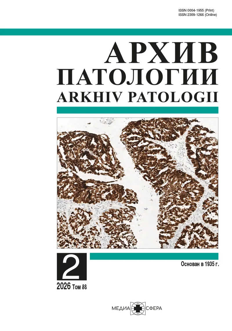
Патоморфологическая картина эозинофильного эзофагита до и после лечения ингибиторами протонной помпы
Резюме
Эозинофильный эзофагит (ЭоЭ) проявляется преимущественно эозинофильной инфильтрацией слизистой оболочки пищевода (более 15 эозинофилов в поле зрения при увеличении в 400 раз). Лечение ингибиторами протонной помпы (ИПП), как правило, является точкой старта терапии у пациентов.
ЦЕЛЬ ИССЛЕДОВАНИЯ
Произвести патоморфологический и морфометрический анализ биоптатов пищевода пациентов с ЭоЭ до и после лечения ИПП.
МАТЕРИАЛ И МЕТОДЫ
Из 84 пациентов с впервые диагностированным ЭоЭ 30 составили группу, получавшую терапию ИПП. Пациентам выполнялась эзофагогастродуоденоскопия с биопсией для первичной диагностики и оценки в динамике. Биоптаты окрашивали гематоксилином и эозином, реактивом Шиффа в сочетании с альциановым синим и по Маллори для выявления фиброза. Морфометрический анализ биоптатов проводился с использованием шкалы EoEHSS для первичной диагностики и EoEHRS для оценки наступления ремиссии.
РЕЗУЛЬТАТЫ
У пациентов с впервые выявленным ЭоЭ пиковое число эозинофилов составило 46 (25—89) в поле зрения при увеличении в 400 раз, частота базально-клеточной гиперплазии — 100%, межклеточного отека — 100%, поверхностного повреждения эпителия — 77,38%, подэпителиального фиброза — 59,52%, эозинофильных абсцессов — 57,14%, поверхностного напластования эозинофилов — 28,57%, дискератоза — 21,43%. Сумма баллов степени по шкале EoEHSS составила 12 (8—13,75), сумма баллов стадии — 10 (8—12). Пиковое число эозинофилов после терапии снизилось во всех наблюдениях (4 (1—12) эозинофила в поле зрения при увеличении в 400 раз), p<0,05. Выраженность и протяженность остальных признаков значительно уменьшилась. Сумма баллов степени по шкале EoEHSS составила 4 (3—8), сумма баллов стадии — 4 (3—7); p<0,05.
ЗАКЛЮЧЕНИЕ
Как для диагностики, так и оценки эффективности терапии ЭоЭ необходимо определять не только пиковое число эозинофилов, но структурные особенности слизистой оболочки пищевода. На терапии ИПП гистологическая ремиссия по критериям EoEHRS достигнута у 40% пациентов.
Ключевые слова
- эозинофильный эзофагит
- ингибиторы протонной помпы
- патоморфологическая шкала оценки эозинофильного эзофагита
- патоморфологическая ремиссия
Дата поступления: 13.08.2025
Дата принятия в печать: 22.12.2025
Дата публикации: 16.04.2026
- Lucendo AJ, Molina-Infante J, Arias Á, von Arnim U, Bredenoord AJ, Bussmann C, Amil Dias J, Bove M, González-Cervera J, Larsson H, et al. Guidelines on eosinophilic esophagitis: evidence-based statements and recommendations for diagnosis and management in children and adults. United European Gastroenterology Journal. 2017;5(3):335-358. https://doi.org/10.1177/2050640616689525
- Dellon ES, Liacouras CA, Molina-Infante J, Furuta GT, Spergel JM, Zevit N, Spechler SJ, Attwood SE, Straumann A, Aceves SS, et al. Updated International Consensus Diagnostic Criteria for Eosinophilic Esophagitis: Proceedings of the AGREE Conference. Gastroenterology. 2018;155(4):1022-1033.e10. https://doi.org/10.1053/j.gastro.2018.07.009
- Кайбышева В.О., Михалева Л.М., Федоров Е.Д. Эозинофильный эзофагит. М.: Медиа Сфера; 2024.
- Molina-Infante J, Gonzalez-Cordero PL, Ferreira-Nossa HC, Mata-Romero P, Lucendo AJ, Arias A. Rising incidence and prevalence of adult eosinophilic esophagitis in midwestern Spain (2007-2016). United European Gastroenterology Journal. 2018;6(1):29-37. https://doi.org/10.1177/2050640617705913
- Robson J, O’Gorman M, McClain A, Mutyala K, Davis C, Barbagelata C, Wheeler J, Firszt R, Smith K, Patel R, Peterson K, Lowichik A, Guthery S. Incidence and Prevalence of Pediatric Eosinophilic Esophagitis in Utah Based on a 5-Year Population-Based Study. Clinical Gastroenterology and Hepatology. 2019;17(1):107-114.e1. https://doi.org/10.1016/j.cgh.2018.06.028
- Arias Á, Lucendo AJ. Incidence and prevalence of eosinophilic oesophagitis increase continiously in adults and children in Central Spain: A 12-year population-based study. Digestive and Liver Diseases. 2019;51(1):55-62. https://doi.org/10.1016/j.dld.2018.07.016
- Arias Á, Lucendo AJ. Epidemiology and risk factors for eosinophilic esophagitis: lessons for clinicians. Expert Review of Gastroenterology and Hepatology. 2020;14(11):1069-1082. https://doi.org/10.1080/17474124.2020.1806054
- Cotton CC, Betancourt R, Randall C, Perjar I, Bookhout C, Woosley JT, Shaheen NJ, Dellon ES. A Model Using Clinical and Endoscopic Characteristics Identifies Patients at Risk for Eosinophilic Esophagitis According to Updated Diagnostic Guidelines. Clinical Gastroenterology and Hepatology. 2021;19(9):1824-1834.e2. https://doi.org/10.1016/j.cgh.2020.06.068
- Lee C, Sievers TJ, Vaughn BP. Diagnosis of Eosinophilic Esophagitis at the Time of Esophageal Food Impaction. Journal of Clinical Medicine. 2023;12(11):3768. https://doi.org/10.3390/jcm12113768
- Hirano I, Moy N, Heckman MG, Thomas CS, Gonsalves N, Achem SR. Endoscopic assessment of the oesophageal features of eosinophilic oesophagitis: validation of a novel classification and grading system. Gut. 2013;62:489-495. https://doi.org/10.1136/gutjnl-2011-301817
- Collins MH, Martin LJ, Alexander ES, Boyd JT, Sheridan R, He H, Pentiuk S, Putnam PE, Abonia JP, Mukkada VA, Franciosi JP, Rothenberg ME. Newly developed and validated eosinophilic esophagitis histology scoring system and evidence that it outperforms peak eosinophil count for disease diagnosis and monitoring. Diseases of Esophagus. 2017;30(3):1-8. https://doi.org/10.1111/dote.12470
- Franciosi JP, Gordon M, Sinopoulou V, Dellon ES, Gupta SK, Reed CC, Gutiérrez-Junquera C, Venkatesh RD, Erwin EA, Egiz A, Elleithy A, Mougey EB. Medical treatment of eosinophilic esophagitis. Cochrane Database of Systematic Reviews. 2023; 7(7):CD004065. https://doi.org/10.1002/14651858.CD004065.pub4
- Maslenkina K, Mikhaleva L, Mikhalev A, Kaibysheva V, Atiakshin D, Motilev E, Buchwalow I, Tiemann M. Assessment of the Severity and the Remission Criteria in Eosinophilic Esophagitis. Biomedicines. 2023;11(12):3204. https://doi.org/10.3390/biomedicines11123204
- Eke R, Li T, White A, Tariq T, Markowitz J, Lenov A. Systematic review of histological remission criteria in eosinophilic esophagitis. JGH Open. 2018;2(4):158-165. https://doi.org/10.1002/jgh3.12059
- Dellon ES, Collins MH, Katzka DA, Mukkada VA, Falk GW, Morey R, Goodwin B, Eisner JD, Lan L, Desai NK, Williams J, Hirano I. ORBIT2/SHP621-302 Investigators. Long-Term Treatment of Eosinophilic Esophagitis With Budesonide Oral Suspension. Clinical Gastroenterology and Hepatology. 2022;20(7):1488-1498.e11. https://doi.org/10.1016/j.cgh.2021.06.020
- Hirano I, Collins MH, Katzka DA, Mukkada VA, Falk GW, Morey R, Desai NK, Lan L, Williams J, Dellon ES. ORBIT1/SHP621-301 Investigators. Budesonide Oral Suspension Improves Outcomes in Patients With Eosinophilic Esophagitis: Results from a Phase 3 Trial. Clinical Gastroenterology and Hepatology. 2022;20(3): 525-534.e10. https://doi.org/10.1016/j.cgh.2021.04.022
- Hirano I, Collins MH, Assouline-Dayan Y, Evans L, Gupta S, Schoepfer AM, Straumann A, Safroneeva E, Grimm M, Smith H, et al. HEROES Study Group. RPC4046, a Monoclonal Antibody Against IL13, Reduces Histologic and Endoscopic Activity in Patients With Eosinophilic Esophagitis. Gastroenterology. 2019;156(3):592-603.e10. https://doi.org/10.1053/j.gastro.2018.10.051
- Collins MH, Martin LJ, Wen T, Abonia JP, Putnam PE, Mukkada VA, Rothenberg ME. Eosinophilic Esophagitis Histology Remission Score: Significant Relations to Measures of Disease Activity and Symptoms. Journal of Pediatric Gastroenterology and Nutrition. 2020;70(5):598-603. https://doi.org/10.1097/MPG.0000000000002637
- Franciosi JP, Mougey EB, Dellon ES, Gutierrez-Junquera C, Fernandez-Fernandez S, Venkatesh RD, Gupta SK. Proton Pump Inhibitor Therapy for Eosinophilic Esophagitis: History, Mechanisms, Efficacy, and Future Directions. Journal of Asthma and Allergy. 2022;15:281-302. https://doi.org/10.2147/JAA.S274524
- Rank MA, Sharaf RN, Furuta GT, Aceves SS, Greenhawt M, Spergel JM, Falck-Ytter YT, Dellon ES. Technical Review on the Management of Eosinophilic Esophagitis: A Report From the AGA Institute and the Joint Task Force on Allergy-Immunology Practice Parameters. Gastroenterology. 2020;158(6):1789-1810.e15. https://doi.org/10.1053/j.gastro.2020.02.039
- Dellon ES, Hirano I. Epidemiology and Natural History of Eosinophilic Esophagitis. Gastroenterology. 2018;154(2):319-332.e3. https://doi.org/10.1053/j.gastro.2017.06.067
- Shaker A. Esophageal remodeling in eosinophilic esophagitis. Current Opinion in Gastroenterology. 2024;40(4):291-298. https://doi.org/10.1097/MOG.0000000000001031
- Arias-González L, Rodríguez-Alcolado L, Laserna-Mendieta EJ, Navarro P, Lucendo AJ, Grueso-Navarro E. Fibrous Remodeling in Eosinophilic Esophagitis: Clinical Facts and Pathophysiological Uncertainties. International Journal of Molecular Sciences. 2024;25(2):927. https://doi.org/10.3390/ijms25020927


Рекомендуем статьи по данной теме:
Фибростенотические осложнения эозинофильного эзофагита.Эндоскопическая хирургия. 2026;32(4):64-68
Диета в терапии эозинофильного эзофагита: серия клинических случаев.Доказательная гастроэнтерология. 2026;15(1):66-75
Развитие сочетанного эозинофильного поражения пищевода и желудка и современные возможности терапии.Доказательная гастроэнтерология. 2025;14(4):94-103
Функциональная диспепсия: подходы к диагностике и терапии в контексте российского опыта.Доказательная гастроэнтерология. 2025;14(2):43-49
Высокоточная детекция эозинофилов на гистологических изображениях при эозинофильном эзофагите с использованием модели машинного обучения YOLO11.Доказательная гастроэнтерология. 2025;14(2):19-29
Ингибиторы протонной помпы: положительные и отрицательные свойства. К 45-летию открытия.Профилактическая медицина. 2024;27(11):135-140
Патология пищевода и бронхиальная астма: патогенетические взаимодействия и возможности оптимизации терапии.Профилактическая медицина. 2024;27(11):129-134