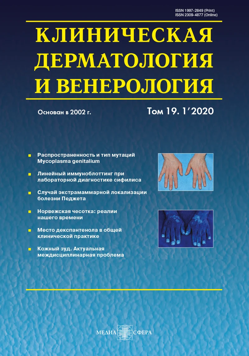
Применение метода визуализации кожи в ультрафиолетовом свете в диагностике заболеваний кожи
Журнал: Клиническая дерматология и венерология. 2020;19(1):43-52.
DOI: 10.17116/klinderma20201901143
Прочитано: 33787 раз
Резюме
Ключевые слова
- неинвазивная диагностика
- визуализация кожи в ультрафиолетовом свете
- лампа Вуда
- нарушения пигментации кожи
- микозы
- бактериальные инфекции кожи
- порфирии
Дата публикации: 30.04.2020
Разработка и совершенствование методов неинвазивной диагностики заболеваний кожи — приоритетное направление развития современной дерматологии. К наиболее распространенным методам неинвазивной диагностики относятся фотографирование, дерматоскопия, УЗИ, оптическая когерентная томография. Одним из простых неинвазивных диагностических тестов является визуализация кожи в УФ-свете (свете Вуда). Однако в российской научной литературе этому методу уделено недостаточно внимания. В статье обобщены сведения о возможностях использования данного метода в практике врача-дерматовенеролога.
Аппаратура
Лампа Вуда. В 1903 г. физик из г. Балтимора Р.У. Вуд сконструировал лампу, представлявшую собой ртутно-кварцевую газоразрядную лампу высокого давления, оборудованную фильтром из силиката бария, содержащим 9% оксида никеля [1]. Такой фильтр позволял задерживать видимый свет и пропускал только длинноволновое УФ-излучение с длиной волны 320—400 нм и пиком эмиссии 365 нм. Впоследствии эта лампа была названа лампой Вуда, а генерируемый свет — светом Вуда. В 1925 г. французские врачи J. Margot и P. Deveze впервые сообщили о возможности использования лампы Вуда в дерматологии [2].
УФ-излучение, индуцируемое лампой Вуда, позволяет визуализировать в коже и волосах флюоресценцию, в основе которой лежит поглощение света электронами и переход их в возбужденное, нестабильное состояние. Возвращение электронов на основную орбиталь сопровождается излучением в видимой области спектра (400—700 нм) в виде специфического свечения. При визуализации кожи в УФ-свете могут наблюдаться различные типы флюоресценции, характер которых зависит от объекта, подвергающегося облучению.
В настоящее время лампа Вуда применяется в повседневной клинической практике (рис. 1) 
Другие источники и устройства. С развитием современных технологий появились новые источники излучения и устройства, позволяющие визуализировать изображения кожи в УФ-свете. В последние годы в качестве источников ультрафиолета все чаще используют люминесцентные лампы со встроенным фильтром с черным покрытием (стекло Вуда), не пропускающим видимый свет. Все большую популярность завоевывают светодиоды, генерирующее излучение аналогичного диапазона.
В МНПЦДК разработано и активно применяется устройство, в котором источники УФ-излучения совмещены с блоком регистрации изображений (рис. 2). 
Применение в клинической практике
Флюоресценция здоровой кожи в УФ-свете обычно слабая или отсутствует, однако при некоторых заболеваниях можно наблюдать интенсивную флюоресценцию очагов поражения с характерными особенностями. Ниже рассмотрены основные аспекты применения метода визуализации кожи в УФ-свете в практике дерматовенеролога (основные показания к применению метода представлены в таблице. 
Нарушения пигментации
При нарушениях пигментации кожи некоторые очаги поражения плохо визуализируются при естественном освещении. Установлено, что одним из основных хромофоров, поглощающих ультрафиолет, является меланин [3]. Уменьшение или увеличение его содержания влияет на характер визуализации кожи в УФ-свете, благодаря чему этот метод можно использовать для неинвазивной диагностики заболеваний, сопровождающихся де-, гипо- и гиперпигментацией кожи.
Де- и гипопигментации. У светлокожих людей очаги де- и гипопигментации кожи нередко не видны при естественном освещении. Осмотр кожи в УФ-свете значительно улучшает их визуализацию. В депигментированных очагах (как правило, при витилиго) меланин в эпидермисе отсутствует или его содержание значительно уменьшено, в результате в коже образуется оптическое окно, позволяющее увидеть флюоресценцию дермального коллагена, индуцируемую ультрафиолетом. При осмотре в УФ-свете очаги депигментации выглядят обычно как ярко светящиеся пятна белого или голубовато-белого цвета с четкими контурами (рис. 3). 
Следует учесть, что изображения кожи в УФ-свете обычно имеют голубоватый оттенок (см. рис. 3б). Для более четкой визуализации светлых и темных фрагментов полученные файлы целесообразно конвертировать в черно-белый формат с расширенным динамическим диапазоном, отображающим оттенки серого цвета (см. рис. 3в).
Визуализация кожи в УФ-свете позволяет не только диагностировать витилиго, но и выявить признаки восстановления пигментации в очагах поражения, а также оценить эффективность лечения (рис. 4). 

При анемическом невусе гипопигментация обусловлена уменьшением содержания в коже гемоглобина, тогда как уровень меланина в ней не изменен. В связи с этим при осмотре в УФ-свете он становится невидимым, поскольку не флюоресцирует.
Гиперпигментации. Визуализация кожи в УФ-свете — хорошее подспорье в диагностике патологии, сопровождающейся гиперпигментацией кожи (хлоазма, веснушки и др.). Известно, что УФ-излучение значительно поглощается гиперпигментированными очагами, тогда как окружающая кожа рассеивает и отражает его. Установлено также, что большая часть УФ-лучей абсорбируется эпидермисом и лишь небольшое количество ультрафиолета достигает дермы. В связи с этим эпидермальная гиперпигментация при осмотре в УФ-свете проявляется более четко, чем при естественном освещении, поскольку контраст между пораженной и здоровой кожей становится более выраженным. При дермальной гиперпигментации различия в окраске пораженной и окружающей здоровой кожи, наоборот, становятся менее заметными. Таким образом, метод визуализации кожи в УФ-свете в определенной мере позволяет оценить глубину преимущественного накопления меланина в коже [3].
Так, например, очаги хлоазмы эпидермального типа при осмотре кожи в УФ-свете выглядят более темными, чем при естественном освещении, и четко контрастируют с окружающей непораженной кожей (рис. 6). 
Следует заметить, что у людей с V и VI фототипами кожи визуализация гиперпигментаций кожи в УФ-свете малоинформативна, поскольку не выявляет контраста между пораженной и здоровой кожей.
Микозы
Дерматофитии. Некоторые виды дерматофитов (грибы рода Microsporum, T.schoenleinii) способны флюоресцировать в УФ-свете. Так, грибы M.canis, M. audouinii, M.distortum, M. ferrugineum при облучении вырабатывают соединение птеридин, флюоресцирующее ярко-зеленым, сине-зеленым или желто-зеленым светом (рис. 7). 
Необходимо подчеркнуть, что выявление флюоресценции дерматофитов не является основанием для постановки диагноза, но позволяет определить локализацию поражения и выбрать пораженные волосы для микроскопического и культурального исследования. Данный метод диагностики можно использовать при проведении профилактических осмотров в детских коллективах (детские сады, школы), а также для оценки эффективности лечения больных. При этом следует иметь в виду, что прекращение свечения в процессе лечения не является основанием для окончания курса терапии.
Разноцветный лишай. Возбудителем разноцветного лишая являются грибы рода Malassezia (Pityrosporum), флюоресцирующие желтовато-белым, золотисто-желтым, медно-оранжевым или буроватым светом. Выявление флюоресценции помогает установить диагноз при субклиническом течении заболевания, а также оценить распространенность поражения и провести дифференциальную диагностику с другой патологией, сопровождающейся нарушением пигментации кожи.
Фолликулит, вызванный грибами Malassezia (Pityrosporum). Грибы Malassezia furfur, способные вызывать воспаление в волосяных фолликулах, при облучении УФ-светом флюоресцируют голубовато-белым светом, что позволяет дифференцировать эту форму фолликулита от бактериального фолликулита.
Бактериальные инфекции кожи
Propionibacterium acnes. Наиболее известными бактериями, вызывающими флюоресценцию в УФ-свете, являются Propionibacterium acnes (P.acnes), входящие в состав бактериальной флоры кожи. Эти бактерии продуцируют копропорфирин III, флюоресцирующий кораллово-красным или оранжево-красным светом. P.acnes играют существенную роль в патогенезе обыкновенных угрей. При осмотре кожи в УФ-свете можно объективно оценить распределение и площадь поражения данной инфекцией. C.W. Choi и соавт. [5] показали, что флюоресценция фолликулов на коже щек коррелирует с количеством P.acnes. Данный метод диагностики может быть полезен не только для определения степени обсемененности кожи P.acnes, но и для оценки эффективности лечения больных при развитии резистентности к антибактериальным препаратам.
Эритразма. Возбудителем эритразмы являются микроорганизмы Corynebacterium minutissimum, флюоресцирующие в УФ-свете кораллово-красным светом (рис. 8). 
Синегнойная инфекция. Синегнойная палочка Pseudomonas aeruginosa является одним из возбудителей гнойно-воспалительных процессов в коже. Она вырабатывает пигмент флюоресцеин, флюоресцирующий при облучении УФ-светом зеленым, желтовато-зеленым или синим светом. Флюоресценция, вызываемая Pseudomonas aeruginosa, используется в клинической практике в качестве теста ранней диагностики инфицирования ожогов и различных ран синегнойной палочкой и помогает подобрать необходимое лечение на ранней стадии заболевания (до получения соответствующей культуры в бактериальном посеве).
Трихомикоз подмышечный (трихобактериоз). Возбудителем заболевания являются бактерии рода Corynebacterium (Corynebacterium flavescens, Corynebacterium sp.), флюоресцирующие в УФ-свете желтым, желто-белым или желтовато-зеленым светом [6].
Порфирии
Порфирии представляют собой группу заболеваний, обусловленных нарушением метаболизма порфиринов. У больных порфириями происходит накопление порфиринов в различных тканях, которое можно выявить в образцах мочи, эритроцитов, кала, тканей зубов и др. При облучении образцов, содержащих порфирины, УФ-светом обычно выявляется характерная флюоресценция красно-розового, розово-красного или оранжево-розового цвета (рис. 9). 
Флюоресценция порфиринов при разных формах порфирий имеет свои особенности. При поздней кожной порфирии флюоресценцию можно обнаружить в моче и биоптатах печени, при вариегатной порфирии порфирины в моче флюоресцируют только в период обострения, тогда как в кале они флюоресцируют даже в период ремиссии [1]. При врожденной эритропоэтической порфирии (болезнь Гюнтера) флюоресценция порфиринов наблюдается в моче, тканях зубов и костном мозге. При эритропоэтической протопорфирии порфирины временно флюоресцируют в эритроцитах (при проведении флюоресцентной микроскопии), в то время как в моче их не выявляют. При острой перемежающейся порфирии флюоресценции обычно не наблюдают.
Другие возможные области применения
Опубликована информация о применении УФ-света для визуализации некоторых других заболеваний, однако обоснованность и эффективность такой диагностики требует дальнейшего изучения. Метод визуализации кожи в УФ-свете используют также для интраоперационной неинвазивной оценки жизнеспособности кожных лоскутов при проведении трансплантации кожи, для визуализации скрытых очагов актинического кератоза и границ новообразований кожи при определении оптимальной тактики хирургического лечения или фотодинамической терапии.
В работе H. Lim и соавт. [7] показана эффективность применения УФ-света в сочетании c использованием экзогенного флюорофора флюоресцеина для интраоперационной неинвазивной оценки жизнеспособности кожных лоскутов при проведении трансплантации кожи. В ряде исследований показана целесообразность применения УФ-света для выявления скрытых очагов актинического кератоза, а также для визуализации границ новообразований кожи (базальноклеточный и плоскоклеточный рак кожи, болезнь Боуэна) с целью определения оптимальной тактики лечения [8—11]. Осмотр в УФ-свете в этих случаях проводят после предварительной аппликации на кожу фотосенсибилизирующего препарата метиламинолевуленовой кислоты, индуцирующей в очагах актинического кератоза и опухолевого роста образование порфиринов (включая протопорфирин IX), флюоресцирующих при облучении красно-розовым светом.
Известны данные о применении УФ-света для выявления атрофических изменений слизистой оболочки влагалища [12], для визуализации субклинических проявлений и оценки распространенности патологического процесса у больных локализованной склеродермией [13], при обследовании очагов порокератоза с целью проведения дифференциальной диагностики с кольцевидной формой красного плоского лишая и кольцевидной гранулемой [14], для диагностики пигментации ногтей, обусловленной применением тетрациклина [15]. Вместе с тем следует иметь в виду, что в ряде случаев при облучении УФ-светом наблюдается ложноположительная флюоресценция, которую могут вызывать некоторые медикаменты (содержащие вазелин, салициловую кислоту и др.), чешуйки, находящиеся на коже, волокна ткани, рубцы, лимонный сок, краска для волос, а также свет, отраженный от белого халата врача, и др. [16].
Заключение
Таким образом, визуализация кожи в УФ-свете — простой и достаточно информативный метод неинвазивной диагностики заболеваний кожи: нарушений пигментации, микозов, бактериальных кожных инфекций, порфирий. В ряде случаев характер свечения может помочь врачу-дерматовенерологу не только диагностировать ту или иную патологию кожи, но и оценить эффективность проводимого лечения. Необходимо проведение дальнейших исследований с целью разработки новых эффективных методов визуализации кожи, основанных на использовании современных технологий.
Авторы заявляют об отсутствии конфликта интересов.
The authors declare no conflict of interest.
Сведения об авторах
Потекаев Н.Н. — https://orcid.org/0000-0002-9578-5490
Кочетков М.А. — https://orcid.org/0000-0002-5788-4666
Волнухин В.А. — https://orcid.org/0000-0002-8363-5188
Петров В.А. — https://orcid.org/0000-0002-9706-1920
Автор, ответственный за переписку: Кочетков М.А. —
e-mail: 1012246@mail.ru


Рекомендуем статьи по данной теме:
Иммунологические перспективы современных методов неинвазивной и малоинвазивной диагностики неалкогольной жировой болезни печени.Адаптивная медицинская иммунология и вопросы общественного здоровья. 2026;2(1):42-59
Возможности применения методов лазерной флюоресцентной спектроскопии и доплеровской флоуметрии при бляшечной склеродермии.Клиническая дерматология и венерология. 2025;24(5):616-624
Скрининг на мутации TERTp методом цифровой капельной ПЦР коллекции из 52 парных образцов ДНК из плазмы крови и опухолевых тканей пациентов с глиобластомой.Вопросы нейрохирургии имени Н.Н. Бурденко. 2025;89(5):87-95