Кавернозная мальформация зрительного нерва: случай из практики и обзор литературы
Журнал: Вопросы нейрохирургии имени Н.Н. Бурденко. 2020;84(1):62-68.
DOI: 10.17116/neiro20208401162
Прочитано: 4037 раз
Резюме
Ключевые слова
- кавернома зрительного нерва
- латеральный супраорбитальный доступ
- «хиазмальная апоплексия»
Дата публикации: 30.04.2020
ЗН — зрительный нерв
КМ — кавернозная мальформация
МРТ — магнитно-резонансная томография
ЦНС — центральная нервная система
Кавернозные мальформации (КМ) зрительных путей являются редкой патологией, составляя примерно 1% от всех каверном центральной нервной системы (ЦНС) [1]. Первое сообщение о КМ зрительного нерва представлено H. Manz и соавт. в 1979 г. [2]. Заболевание обычно проявляется «хиазмальной апоплексией» в виде острого развития зрительных нарушений, головной и ретроорбитальной боли, а также тошноты [3]. Часто пароксизмальной симптоматике за несколько недель или месяцев предшествуют преходящее снижение остроты зрения и умеренные головные боли. Симптомы могут оставаться на одном уровне и не прогрессировать, однако у некоторых больных возможно необратимое повреждение зрительных нервов (ЗН) на фоне кровоизлияния.
Хирургическое лечение КМ выполняется с целью декомпрессии ЗН и исключения повторных кровоизлияний. Выбор доступа основывается на размерах и локализации КМ, данных нейровизуализации и клинической симптоматике. Традиционно используются птериональный, латеральный супраорбитальный, супраорбитальный трансбровный доступы [4—8]. Тотальное удаление КМ — основная задача микрохирургического лечения. Частичное удаление может грозить повторными кровоизлияниями с последующим ухудшением зрения [9, 10].
Мы представляем случай из практики — клинический пример удаления КМ левого ЗН.
Клинический случай
Пациентка К., 32 лет, обратилась в нейрохирургическое отделение с жалобами на периодические головные боли и снижение зрения на левом глазу. Из анамнеза известно, что за 2 нед до госпитализации больную начали беспокоить периодические головные боли с последующим резким падением остроты зрения слева. Амбулаторно выполнена магнитно-резонансная томография (МРТ) головного мозга, выявлено новообразование левого З.Н. Пациентка обратилась для оперативного лечения.
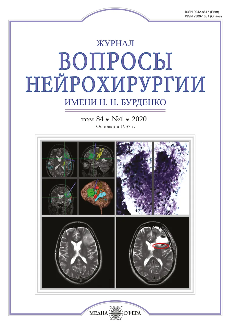
При МРТ головного мозга определено объемное образование левого ЗН в интракраниальном сегменте с повышенным МР-сигналом в режимах Т2 и DWI, размерами 9,5×11,1 мм, округлой формы с четкими контурами, не накапливающее контрастный препарат (рис. 1, а—г). 
При осмотре офтальмологом выявлены снижение остроты зрения слева до 0,3 и правосторонняя гемианопсия с преобладанием выпадения поля зрения в левом глазу. Рефракция левого глаза не отличалась от таковой противоположного глаза. Глазодвигательных нарушений не было. При осмотре глазного дна выявлено, что диск ЗН бледно-розовый, не отечен, с четкими границами.
Учитывая жалобы, анамнестические данные, клиническую симптоматику и данные МРТ, было принято решение о микрохирургическом удалении образования из латерального супраорбитального доступа.
После выполнения краниотомии вскрыта твердая мозговая оболочка, осуществлен доступ к базальным цистернам для релаксации мозга. Фиксированная тракция лобной доли не использовалась. Визуализирован увеличенный в размере левый ЗН желтого цвета с обеднением сосудистого рисунка. Под пиальной оболочкой нерва визуализировано объемное новообразование вишневого цвета, напоминающее каверному, с признаками кровоизлияния и компрессией нерва; после рассечения арахноидальных спаек нерв оставался малоподвижным, что затрудняло оценку. С целью мобилизации и улучшения визуализации выполнена декомпрессия ЗН путем частичной резекции верхней стенки зрительного канала. При удалении образования выявлены очаги кровоизлияний с плотными сгустками крови и небольшое количество однородных коричнево-красноватых масс полужидкой консистенции в подостро-хронической стадии, легко отделяемых от З.Н. После декомпрессии новообразование отделено от ЗН и удалено тотально, отправлено на гистологическое исследование. Закрытие раны послойное, костный лоскут установлен на место и фиксирован титановыми минипластинами, наложен непрерывный внутрикожный шов. Дренаж не использовался. Осложнений не отмечено. Этапы удаления представлены на рис. 2. 

Больная активизирована в 1-е сутки после операции. При осмотре офтальмологом на 4-е сутки выявлено незначительное ухудшение остроты зрения до 0,25 и небольшое увеличение площади выпавшего поля зрения слева. Выписана на 5-е сутки. При морфологическом исследовании выявлены сгустки крови в стадии организации, стенка новообразования представлена соединительной тканью и мелкими очагами кровоизлияний; результат исследования — КМ.
При осмотре через 3 мес после операции отмечены восстановление остроты зрения слева до 0,3 и регресс гемианопсии до дооперационного уровня; на изображениях контрольных МРТ головного мозга — послеоперационные изменения в области левого З.Н. Данных за повторные кровоизлияния и рецидив каверномы нет (рис. 4). 
Обсуждение
Частота выявления КМ черепно-мозговых нервов достаточно мала. В литературе известны случаи поражения II—V, VII—VIII пар черепно-мозговых нервов [5, 11]. К настоящему времени известно около 50 сообщений о КМ зрительных путей [12, 13]. В большинстве наблюдений клиническая симптоматика проявляется во 2—4-й декадах жизни [14]. Как правило, нет зависимости наличия КМ от пола, однако имеются наблюдения о преобладании кровоизлияний из КМ у женщин [15]. В клинической картине КМ ЗН преобладают зрительные нарушения. «Хиазмальная апоплексия» обусловлена кровоизлиянием в КМ [16]. По данным литературы, частота кровоизлияний в КМ составляет от 0,7 до 3,1% в год [14, 17]. Первичное кровоизлияние повышает риск последующих кровоизлияний, однако при динамическом наблюдении риск через 2—3 года постепенно снижается [18]. В литературе отсутствуют убедительные статистические данные о частоте кровоизлияний при КМ в области зрительных путей, однако считается, что подобный риск выше, чем для остальных отделов ЦНС [19, 20].
Ведущая роль в диагностике КМ принадлежит МРТ. Нейровизуализационная характеристика КМ зрительных путей схожа с КМ остальных отделов ЦНС [19, 21, 22]. Используя компьютерную томографию в качестве диагностики КМ ЗН, следует проводить дифференциальный диагноз с опухолью и тромбированной аневризмой, так как гиперденсивные участки и возможные кальцификаты могут встречаться и при описанной ранее патологии [23]. Церебральная ангиография позволяет исключить наличие патологических сосудов артериовенозной мальформации, однако при верифицированной КМ возможны и ангиографические находки венозных ангиом [1, 24]. Помимо этого проводится дифференциальный диагноз с глиомами и менингиомами зрительных нервов, краниофарингиомами (характерным является накопление контрастного препарата), апоплексией гипофиза, невритом зрительного нерва [25—30].
По мнению многих авторов, микрохирургическое лечение является методом выбора для КМ зрительных путей. Тотальная резекция КМ — единственный способ предотвращения повторных кровоизлияний, так как любая резидуальная часть КМ может быть их источником [5, 6, 19—21].
В большинстве случаев показано раннее хирургическое лечение КМ ЗН [5, 6, 21]. У пациентов с «хиазмальной апоплексией» предпочтительны установление диагноза и проведение оперативного вмешательства в течение 24 ч после появления симптоматики с целью быстрого устранения компрессии зрительных путей [19]. Своевременная диагностика важна, поскольку даже при полной потере зрения может произойти его восстановление в случае быстрого выполнения декомпрессии зрительных нервов [31].
Основными особенностями микрохирургического этапа являются резекция крыши канала ЗН для мобилизации и декомпрессии нерва; вскрытие пиальной оболочки нерва с продольной диссекцией; выделение и резекция КМ острым путем. Такой подход позволяет сохранить имеющееся зрение и создать условия для его восстановления.
Применение стереотаксической радиохирургии в лечении КМ больших полушарий и ствола головного мозга имеет определенный успех, однако использование этой методики при локализации КМ в передних отделах зрительных путей сопряжено с высоким риском постлучевой нейропатии зрительного нерва [32].
Работа J. Liu и соавт. является наиболее крупным обзором 65 случаев КМ зрительных путей и гипоталамуса в литературе. Авторы сообщают, что в 60% случаев КМ удалены тотально, субтотальная резекция выполнена у 4 (6%) пациентов, биопсия — у 4 (6%), биопсия с декомпрессией зрительных путей — у 15 (23%). Улучшение зрения отмечено у большинства (75%) пациентов, у 19,3% — изменений не отмечено, у 2 (4%) пациентов в послеоперационном периоде зрение ухудшилось. Консервативную терапию получали 2 пациента: у одного больного отмечено улучшение зрения, у второго — ухудшение. В 1 наблюдении применялось радиохирургическое лечение, но без выраженного эффекта в отношении зрительных функций [12].
В отечественной литературе заслуживает внимания работа А.Н. Шкарубо с соавт., представляющими случай каверномы хиазмы у 23-летней пациентки, у которой помимо зрительных нарушений имелись эндокринные расстройства в виде полиурии, полидипсии, в связи с чем дифференциальный диагноз проводили между краниофарингиомой и глиомой хиазмы. Капсула гематомы и сгустки крови удалены, гистологически подтверждена КМ [33].
Еще одна работа, посвященная интрахиазмальным гематомам, опубликована Ю.А. Григоряном и соавт. В ней представлен случай «хиазмальной апоплексии» у пациентки 41 года. Выполнена операция из птерионального доступа с удалением интрахиазмальной гематомы. Авторами проведен анализ публикаций по теме, касающейся особенностей диагностики и лечения интрахиазмальных кровоизлияний [34].
Заключение
КМ ЗН — достаточно редкая и сложная патология как в плане верификации диагноза, так и для микрохирургического лечения. В случае проявления «хиазмальной апоплексии» раннее хирургическое вмешательство является методом выбора. Тотальное удаление КМ может способствовать улучшению зрительных функций.
Участие авторов
Концепция и дизайн исследования — Р.Д., О.Д., В.Л.
Сбор и обработка материала — Р.Д., А.П., Э.С.
Статистическая обработка — А.П., Р.К.
Написание текста — Р.Д., А.П.
Редактирование — В.Л., О.Д.
Авторы заявляют об отсутствии конфликта интересов.
The authors declare no conflicts of interest.
Сведения об авторах
Джинджихадзе Р.С. — https://orcid.org/0000-0003-3283-9524
Древаль О.Н.— https://orcid.org/0000-0002-8944-9837
Лазарев В.А. — https://orcid.org/0000-0001-9663-0960
Поляков А.В. — https://orcid.org/0000-0001-7413-1968
Камбиев Р.Л. — https://orcid.org/0000-0003-3694-4698
Салямова Э.И. — https://orcid.org/0000-0001-6449-7114
Автор, ответственный за переписку: Джинджихадзе Р.С. — e-mail: brainsurg77@gmail.com
Комментарий
Работа представляет собой описание единичного наблюдения, но это не умаляет ее ценности ввиду того, что она затрагивает крайне актуальную проблему — дифференциальный диагноз патологических образований хиазмально-селлярной области. Несмотря на то что порой ошибочный предварительный диагноз не влияет на выбор хирургического доступа к новообразованию, интраоперационная находка такого плана способна поставить в тупик даже достаточно опытного хирурга.
Безусловно, принципиально важной является своевременная диагностика. Осмотр нейроофтальмолога, МРТ, выполненная в кратчайшие сроки от начала заболевания, — залог оптимального результата лечения. При этом качество обследования также имеет большое значение. Следует помнить, что в подобной ситуации необходимо обязательно дополнить исследование последовательностью SWAN.
Авторы подробно описывают хирургическую технику и делают очень важный акцент на мобильности зрительного нерва в ходе манипуляций. Здесь необходимо добавить, что, в зависимости от конкретной ситуации (в том числе, от распространенности кавернозной мальформации), можно как не выполнять декомпрессию зрительного нерва вовсе, так и выполнять ее в расширенном виде с клиноидэктомией.
Вероятность столкнуться с подобной патологией невелика, но знание деталей дооперационной картины и хирургической техники позволит значительно повысить шансы пациента на благоприятный исход, что делает данную работу весьма полезной для практикующего нейрохирурга.
В.А. Черекаев, Н.В. Ласунин (Москва)


Рекомендуем статьи по данной теме:
Дерматоскопические и патогистологические особенности дерматофибром.Клиническая дерматология и венерология. 2026;25(4):685-694
Высокая безопасность филлеров на основе агарозы. Собственное наблюдение.Клиническая дерматология и венерология. 2026;25(4):676-684
Коррекция признаков старения кожи лица с помощью экзосом.Клиническая дерматология и венерология. 2026;25(4):668-674
Антибактериальная резистентность в дерматологии: современный взгляд на проблему и место амоксициллина и клавулановой кислоты в лечении бактериальных инфекций кожи.Клиническая дерматология и венерология. 2026;25(4):660-667
Дерматозы как ятрогенное осложнение инвазивных аппаратных процедур.Клиническая дерматология и венерология. 2026;25(4):652-658
Эволюция эмолентов и активных компонентов в дерматологии.Клиническая дерматология и венерология. 2026;25(4):641-650
Лекарственно-индуцированная саркоидоподобная реакция на фоне терапии ингибиторами BRAF/MEK метастатической меланомы.Клиническая дерматология и венерология. 2026;25(4):632-640
Мазь такролимуса: селективный T-клеточный иммуносупрессант в дерматологии.Клиническая дерматология и венерология. 2026;25(4):619-630
Клинический случай лечения препаратом нетакимаб пациента с псориазом среднетяжелой степени тяжести, ассоциированным с метаболическим синдромом.Клиническая дерматология и венерология. 2026;25(4):613-618
Современные подходы к терапии NCMT-инфекций, передаваемых половым путем (N. gonorrhoeae, C. trachomatis, M. genitalium, T. vaginalis). Часть II. M. genitalium.Клиническая дерматология и венерология. 2026;25(4):603-612