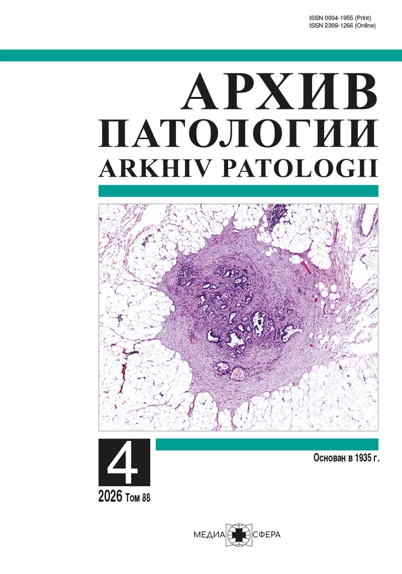
Внелегочный лимфангиолейомиоматоз с поражением забрюшинных лимфатических узлов
Резюме
Лимфангиолейомиоматоз (ЛАМ) — редкое заболевание неясной этиологии, которое встречается почти исключительно у лиц женского пола, преимущественно репродуктивного возраста. Заболевание характеризуется пролиферацией гладкомышечных клеток чаще всего легких, однако существуют случаи развития экстрапульмонального ЛАМ, например, в лимфатических узлах средостения, брюшной полости и забрюшинного пространства. Клиническое проявление ЛАМ тазовых лимфатических узлов имеет скрытый характер: проявляется в виде симптомов сдавления при большом объеме опухоли, но чаще всего диагностируется случайно в материале лимфатических узлов, удаленных во время гинекологических онкологических операций. В статье приведены данные литературы и собственного наблюдения ЛАМ лимфатических узлов у пациентки с метастатическим раком эндометрия.
Ключевые слова
- лимфангиолейомиоматоз
- ЛАМ
Дата поступления: 26.02.2026
Дата принятия в печать: 24.04.2026
Дата публикации: 30.07.2026
- Rabban JT, Firetag B, Sangoi AR, Post MD, Zaloudek CJ. Incidental Pelvic and Para-aortic Lymph Node Lymphangioleiomyomatosis Detected During Surgical Staging of Pelvic Cancer in Women Without Symptomatic Pulmonary Lymphangioleiomyomatosis or Tuberous Sclerosis Complex. The American journal of surgical pathology. 2015;39(8): 1015-1025. https://doi.org/10.1097/PAS.0000000000000416
- McCormack FX, Travis WD, Colby TV, Henske EP, Moss J. Lymphangioleiomyomatosis: calling it what it is: a low-grade, destructive, metastasizing neoplasm. American journal of respiratory and critical care medicine. 2012;186(12): 1210-1212. https://doi.org/10.1164/rccm.201205-0848OE
- Travis WD, Colby TV, Corrin B, Shumosato Y, Brambilla E; in collaboration with L. H. Sobin and pathologists from fourteen countries. Histological typing of lung and pleural tumors. Berlin: Springer; 1999.
- Travis WD, Brambilla E, Muller-Hermlink HK, Harris CC. Pathology and genetics: tumours of the lung, pleura, thymus and heart. Lyon: IARC; 2004.
- WHO Classification of Tumours Editorial Board. Thoracic tumours 5th ed.; vol. 5. Lyon (France): IARC;2021.
- WHO Classification of Tumours Editorial Board. Haematolymphoid tumours 5th ed.; vol. 11. Lyon (France): IARC; 2024.
- Matsui K, Tatsuguchi A, Valencia J, Yu ZX, Bechtle J, Beasley MB, Avila N, Travis WD, Moss J, Ferrans VJ. Extrapulmonary lymphangioleiomyomatosis (LAM): clinicopathologic features in 22 cases. Human pathology. 2000;31(10): 1242-1248. https://doi.org/10.1053/hupa.2000.18500
- Ando H, Ogawa M, Watanabe Y, Tsurunaga K, Nakamura C, Tamura H, Ikeda K, Takahashi R, Nakajima R, Yoshida S, Tsujie T, Doi R, Wakimoto A, Adachi S. Lymphangioleiomyoma of the Uterus and Pelvic Lymph Nodes: A Report of 3 Cases, Including the Potentially Earliest Manifestation of Extrapulmonary Lymphangioleiomyomatosis. International journal of gynecological pathology: official journal of the International Society of Gynecological Pathologists. 2020;39(3): 227-232. https://doi.org/10.1097/PGP.0000000000000589
- Schoolmeester JK, Park K J. Incidental Nodal Lymphangioleiomyomatosis Is Not a Harbinger of Pulmonary Lymphangioleiomyomatosis: A Study of 19 Cases With Evaluation of Diagnostic Immunohistochemistry. The American journal of surgical pathology. 2015;39(10): 1404-1410. https://doi.org/10.1097/PAS.0000000000000470
- Iwasa Y, Tachibana M, Ito H, Iwami S, Yagi H, Yamada S, Okagaki A, Ban C, Mano M, Kodama Y, Ueda M. Extrapulmonary lymphangioleiomyomatosis in pelvic and paraaortic lymph nodes associated with uterine cancer: a report of 3 cases. International journal of gynecological pathology: official journal of the International Society of Gynecological Pathologists. 2011;30(5): 470-475. https://doi.org/10.1097/PGP.0b013e318212e1e6
- Xiao S, Chen Y, Tang Q, Xu L, Zhao L, Wang Z, Yu E. Pelvic Lymph Node Lymphangiomyomatosis Found During Surgery for Gynecological Fallopian Tube Cancer: A Case Report and Literature Review. Frontiers in medicine. 2022;9: 917628. https://doi.org/10.3389/fmed.2022.917628
- Nagasaka T, Murakami Y, Sasaki E, Hosoda W, Nakanishi T, Yatabe Y. Minute perivascular epithelioid cell (PEC) nests in the abdominal lymph nodes--a putative precursor of PEComa. Pathology international. 2015;65(4): 193-196. https://doi.org/10.1111/pin.12262
- Blausteins pathology of the female genital tract. 6th ed. New York: Springer; 2011. https://doi.org/10.1007/978-1-4419-0489-8.
- Flavin RJ, Cook J, Fiorentino M, Bailey D, Brown M, Loda MF. β-Catenin is a useful adjunct immunohistochemical marker for the diagnosis of pulmonary lymphangioleiomyomatosis. American journal of clinical pathology. 2011;135(5): 776-782. https://doi.org/10.1309/AJCPPC9EX1ZHMRMA
- Matsui K, Travis WD, Gonzalez R, Terzian JA, Rosai J, Moss J, Ferrans VJ. Association of lymphangioleiomyomatosis (LAM) with endosalpingiosis in the retroperitoneal lymph nodes: report of two cases. International journal of surgical pathology. 2001;9(2): 155-162. https://doi.org/10.1177/106689690100900212
- Corrin B, Liebow AA, Friedman PJ. Pulmonary lymphangiomyomatosis. A review. The American journal of pathology. 1975;79(2): 348-382.
- Ho TB, Hull JH, Hughes NC. An 86-year-old female with lymphangioleiomyomatosis. The European respiratory journa. 2006;28(5) 1065. https://doi.org/10.1183/09031936.00076506
- Kebria M, Black D, Borelli C, Modica I, Hensley M, Chi DS. Primary retroperitoneal lymphangioleiomyomatosis in a postmenopausal woman: a case report and review of the literature. International journal of gynecological cancer : official journal of the International Gynecological Cancer Society. 2007;17(2): 528-532. https://doi.org/10.1111/j.1525-1438.2007.00785.x
- Закурдаев Е.И., Багателия З.А., Чижиков Н.П. и др. Случай лимфангиолейомиоматоза тазовых лимфатических узлов при раке эндометрия с подозрением на регионарные метастазы. Онкопатология 2024;7(4):33-5. https://doi.org/10.17650/2618-7019-2024-7-4-33-35


Рекомендуем статьи по данной теме:
Лимфангиолейомиоматоз забрюшинных лимфатических узлов: опухоль с неоднозначным клиническим течением.Архив патологии. 2025;87(5):54-59
Современные подходы к диагностике кистозных заболеваний легких.Респираторная медицина. 2025;1(1):30-37